X-ray and CT can help clinicians determine whether it is safe for patients with embedded bullet fragments to undergo MRI for other indications such as brain cancer, spinal cord injury, or joint disorders, according to a study published December 29 in the American Journal of Roetgenology.
The results could allow more patients with these fragments to make use of MRI if needed, wrote a team led by Dr. Arthur Fountain of Emory University in Atlanta.
"Patients with ballistic embedded fragments are frequently denied MRI because the bullet composition cannot be determined without shell casings," the team wrote. "We found that radiography and CT can be used to identify nonferromagnetic projectiles that are safe for MRI."
Embedded ferromagnetic objects can cause injury on MRI due to migration, torque, or heating effects -- especially if they are in soft tissues near vascular or neural structures, the team noted. But the group found that current information on the safety of imaging patients with embedded bullet fragments with MRI is too general, and it can be difficult to identify a bullet's composition before imaging.
"The firearm, bullet, or spent case is frequently not available for identification either in the acute setting or, more commonly, during imaging for an unrelated health issue at a later date," Fountain and colleagues wrote. "The purpose of our study was to determine whether the radiographic and CT appearance of ballistic projectiles predicts their composition and to characterize ... the effects of a 1.5-tesla MRI magnetic field on representative bullets."
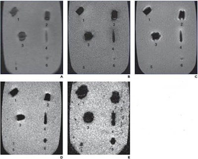
The study involved firing commercially available handgun and shotgun bullets into ballistic gelatin and imaging the gelatin using x-ray and CT. The team then used 1.5-tesla MRI T1 and T2 weighted sequences to visualize unfired bullets suspended in different gelatin blocks, assessing magnetic attraction, torque, and heating effects of the metal items in the gelatin.
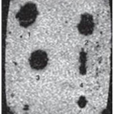
 MRI of nonferromagnetic ballistics suspended in gelatin. Scout (A), T1-weighted spin-echo; (B), T2-weighted SE (C); T2-weighted gradient-recalled echo; (D), and T2-weighted gradient-recalled echo; (E) MR images show jacket hollow point .45 automatic Colt pistol bullet (1), solid lead .45 Long Colt bullet (2), full metal jacket automatic Colt pistol bullet (3), 5.56-mm full metal jacket bullet (4), #7 lead shotgun pellet (5), and 5-mm lead air gun pellet (6). On all sequences, metallic artifact is minimal. Images and caption courtesy of the American Roentgen Ray Society (ARRS).
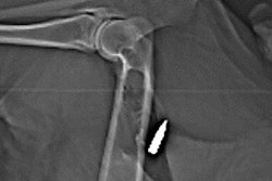
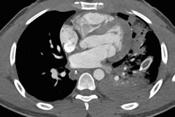
X-ray and CT images helped determine bullet fragment composition by identifying the presence or absence of a debris trail. They also indicated that ferromagnetic bullets showed mild torque forces and imaging artifacts, while nonferromagnetic bullets did not show these effects. None of the bullets visualized on MRI heated the gelatin higher than the U.S. Food and Drug Administration limit of 35.6○ Fahrenheit.
"The bullets we tested that left a debris track or appeared deformed on radiographs or CT images are not ferromagnetic, do not significantly heat up or produce detectable force or torque during MRI, and do not produce much artifact on T1- or T2-weighted MR images," the authors wrote.
The study suggests that x-ray and CT could make MRI more available in patients who need MRI for other indications but also have embedded bullet fragments, according to the team.
"Nonferromagnetic ballistic projectiles do not undergo movement or heating during MRI, and the imaging modality can be performed when medically necessary without undue risk and with limited artifact susceptibility on the resulting images, even when the projectile is in or near a vital structure," the investigators concluded.


