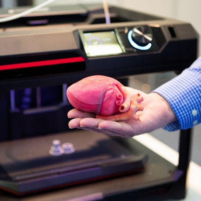
3D printing may be able to help in stratifying risk and making surgical decisions for patients with anomalous aortic origin of the coronary arteries (AAOCA), one of the most common causes of sudden cardiac death in children, according to research published online January 21 in Computer Methods and Programs in Biomedicine.
In a small retrospective feasibility study, researchers led by Jayanthi Parthasarathy, PhD, of the Ohio State University College of Medicine found that they could accurately produce 3D-printed models from CT angiography (CTA) studies in these patients.
"[3D-printed] models derived from advanced imaging can accurately represent patient-specific pathologic anatomy of children with AAOCA, facilitating downstream hemodynamic flow evaluation and refinement of criteria for risk stratification, patient education, and therapeutic decision-making," the authors wrote.
AAOCA is a congenital abnormality that isn't well understood. Although several morphological abnormalities have been identified by advanced imaging, the mechanisms leading to ischemia and/or sudden cardiac death are unknown, according to the researchers, which included co-senior authors Lakshmi Prasad Dasi, PhD, of Georgia Institute of Technology in Atlanta, and Dr. Rajesh Krishnamurthy of Nationwide Children's Hospital in Columbus, OH.
"Currently, there is no definitive information on whether exercise restriction or surgical intervention improves the risk of [sudden cardiac death] on patients with AAOCA," the authors wrote. "[A] critical barrier to progress is a lack of understanding of the exact mechanism leading to ischemia and/or [sudden cardiac death] in these patients and the subsequent inability to perform accurate risk stratification and decide management."
As a result, the researchers sought to assess the accuracy and feasibility of 3D-printed models derived from CTA exams in eight patients from Nationwide Children's Hospital from 2018 to 2019. Using a Connex 350 printer (Stratasys), they created 10 models with an aorta wall thickness of 2 mm and a coronary artery wall thickness of 1.5 mm. Two of the models were produced after surgery.
The models were printed using Agilus30, a flexible photopolymer that's coated with a thin layer of parylene, polyurethane, and silicone. At 2 mm thickness, the dynamic modulus of Agilus30 was found to be close to native aortic tissue, according to the researchers.
Next, CT exams were acquired on the 3D-printed models to assess the accuracy of their morphological characteristics. Two pediatric cardiac radiologists with more than 15 years of experience then provided a blinded review of the CT exams of the 3D models and the patient's original CTA exams.
After performing Pearson's correlation analysis, the researchers found a high level of concordance between the 3D-printed models and the patient's CTA exam:
- Aorta (linear): r = 0.9959
- Aorta (cross-sectional, dimensions): r = 0.9949
- Coronaries (linear): r = 0.9538
- Coronaries (cross-sectional, dimensions): r = 0.9538
What's more, the mean difference in the surface contour map was 0.08 ± 0.29 mm.
"Geometrically accurate AAOCA models preserving morphological characteristics, essential for risk stratification and decision-making, can be 3D printed from a patient's CTA," the authors wrote.
These models could then be used in blood-flow simulation studies for risk stratification by calculating fractional flow reserve (FFR) and pressure during rest and exercise, they said.
"We hope that the significance and impact of our research will extend beyond the AAOCA community, since such patient-specific flow-modeling approaches have widespread applicability in congenital heart diseases, which often have a biomechanical basis for poor outcomes before and after palliation," the researchers wrote.


