Virtual surgical planning and 3D-printed models can yield lower complication rates in maxillectomy reconstructions, according to research from the Mayo Clinic published online April 1 in JAMA Otolaryngology -- Head and Neck Surgery.
In a retrospective study, researchers led by Dr. Eric Moore found that a group of patients who had received virtual surgical planning with 3D-printed models for maxillectomies that required microvascular reconstruction had a much lower rate of lateral rhinotomy than a cohort of similar patients treated prior to adoption of these methods.
"Preoperative [virtual surgical planning], use of 3D printed models and cutting guides, and intraoperative fixation of the reconstruction to the model may eliminate the use of [lateral rhinotomy] for even extensive maxillectomy reconstruction," they wrote. "Avoidance of [lateral rhinotomy] may decrease the rate of maxillectomy-related complications."
Although a transoral approach can be used to perform maxillectomy, this technique can be challenging, according to the researchers. As a result, they sought to assess the benefit of virtual surgical planning and 3D models in patients undergoing total and partial maxillectomy reconstruction.
They retrospectively reviewed 38 patients who had undergone maxillectomy with free-flap reconstruction at their tertiary academic medical center between January 1, 2008, and October 3, 2019. Of the 38 patients, 15 received maxillectomy without virtual surgical planning, while 23 underwent the same procedure but after the institution adopted virtual surgical planning and the use of 3D-printed models.
With this process, surgical planning meetings are held with the radiologist to review high-resolution CT images of the maxilla and the donor site for the free flap. After the surgeon and radiologist discuss the reconstruction goals, the radiologist transfers the images to the onsite anatomical modeling laboratory. Next, the images are imported into the Mimics software (Materialise) and segmented in order to separate the different anatomical structures.
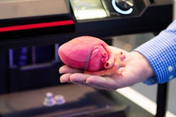
Created from these segmentations, a virtual 3D anatomical model containing several parts is exported into a stereolithographic file. Finally, Materalise's 3-Matic software is utilized for any needed postprocessing before the 3D model is printed using various liquid polymers on a PolyJet Connex 350 3D printer (Stratasys).
In a similar process to the 3D model, cutting guides are produced to enable precise placement and orientation of the osteotomies on the maxilla, as well as the bone of the reconstructive flap, according to the researchers.
"These cutting guides are an integral part of the planning and execution to allow for the avoidance of lateral rhinotomy," the authors wrote.
The sterilized model and the cutting guides are brought into the operating room.
| Benefits of virtual surgical planning and 3D models in maxillectomies | ||
| Before adoption of virtual surgical planning | After adoption of virtual surgical planning | |
| Patients who needed lateral rhinotomy | 12 (80%) | 1 (4%) |
| Patients who had lateral rhinotomy complications | 6 (50%) | 0 |
In other results, the researchers noted that there were no flap failures in the group of patients who received lateral rhinotomy, but only one flap failure among patients who did not undergo the procedure.
"This study suggests that [virtual surgical planning] may help with planning, execution, placement, and fixation for complex free flap maxillectomy defect reconstruction through a transoral minimally invasive approach," the authors wrote.


