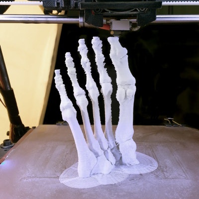
CT images acquired at ultralow radiation doses can be used to produce 3D-printed models of wrist fractures that are just as useful as 3D models generated from standard-dose CT exams, according to research published online December 23 in the European Journal of Radiology.
A team of Chinese researchers led by Mengqiang Xiao of Guangdong Hospital of Traditional Chinese Medicine in Zhuhai City found that an ultralow-dose CT protocol yielded 97% lower radiation dose and enabled production of 3D-printed models that were deemed equivalent to those manufactured from standard-dose images for distal radial fractures.
"Therefore, this protocol can meet the needs of 3D printing models for preoperative assessments," the authors wrote.
Although the effects of low-dose CT techniques on 3D printing have been unknown, the researchers hypothesized that low-dose image acquisition could be sufficient for the creation of 3D models of distal radial fractures. To test this hypothesis, they enrolled 76 consecutive patients with clinical suspicion of distal radial fractures and randomly divided them into groups that received unenhanced CT scans at either standard-dose (120 kV and 100 mA) or ultralow-dose (80 kV and 10 mA) protocols on an Aquilion One CT scanner (Canon Medical Systems).
Canon's adaptive iterative dose reduction (AIDR) 3D adaptive iterative reconstruction software was used to reconstruct all of the images. 3D-printed models were then generated from the reconstructed 3D CT data at 0.5-mm slice intervals.
After an experienced musculoskeletal radiologist objectively measured image-quality parameters, two other experienced musculoskeletal radiologists rated the image quality of the fracture line on all images using a three-point scale: 3 = good (good visualization of the fracture line and excellent definition of the distal radial fracture); 2 = adequate (adequate visualization of the fracture line, slightly affected by image noise, as well as good visualization of the distal radial fracture); and 1 = poor (inadequate visualization of the fracture line and poor definition of the distal radial fracture).
Next, two experienced orthopedic surgeons evaluated the 3D-printed models on a three-point scale: 3 = good (bone model surface is regular and flat, appropriate for preoperative assessment); 2 = adequate (slightly irregular surface, appropriate for preoperative assessment without influence from minor model impairment); and 1 = poor (irregular surface, not appropriate for preoperative assessment).
To measure radiation dose, the researchers used output provided automatically by the scanner for volume CT dose index (CTDIvol) and dose-length product (DLP). DLP was multiplied by the conversion coefficient k to obtain the effective radiation dose for each patient.
| Standard-dose vs. ultralow dose CT for 3D-printed models of distal radial fractures | ||
| Standard-dose CT scans | Ultralow-dose CT scans | |
| Image quality scores | ||
| Mean image quality score (1-3) by musculoskeletal radiologists of fracture line | 2.92 ± 0.27 | 2.16 ± 0.37 |
| Mean quality score (1-3) by orthopedic surgeons of 3D-printed models | 3 | 3 |
| Radiation dose | ||
| DLP | 48.7 mGy x cm | 1.4 mGy x cm |
| CTDIvol | 4.3 mGy | 0.1 mGy |
| Effective dose | 9.75 µSv | 0.28 µSv |
The difference in the objective image-quality parameters and the image-quality scores for the fracture line were statistically significant (p < 0.001). However, both standard-dose and ultralow-dose images were considered to have adequate diagnostic performance, and the researchers found no difference in the subjective scores of 3D-printed models.
Although diagnostic performance wasn't affected by the use of the ultralow-dose protocol, the investigators noted that the quality of CT images and 3D-printed models were still lower than standard-dose images.
"Nevertheless, ultra-low-dose 3D printing models still showed its unique advantages for preoperative evaluation and doctor-patient communication," the authors wrote. "Therefore, ultra-low-dose scan protocol in 3D printing models could be beneficial to both patients and orthopedic surgeons."


