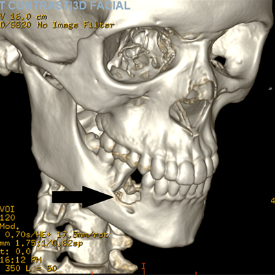
Imaging and lab work helped diagnose a central giant cell granuloma (CGCG) -- a rare, benign, aggressive tumor -- in a 17-year-old with jaw pain and swelling. The case was published on September 26 in the Cureus Journal of Medical Science.
A CT scan and histopathology results revealed a central giant cell granuloma centered in the right lower jaw with erosion of about 2.2 cm through the inner and outer cortex of the jaw. The patient made a full recovery after oral surgery, the authors wrote.
"Early diagnosis, prompt intervention, and complete removal of CGCG lesions result in an excellent prognosis and low recurrence rates," wrote the group, led by Dr. Gurleen Kaur Kahlon from the Brooklyn Hospital Center in New York City.
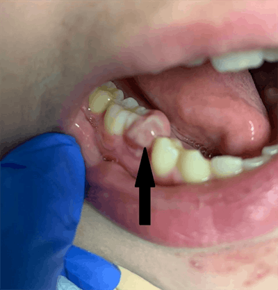 An oral exam showed gingival swelling. Images courtesy of Kahlon et al. Licensed under CC BY 4.0.
Common symptoms, many possible diagnoses
Visiting the emergency department for jaw pain associated with swelling is not unusual. These symptoms can be associated with numerous conditions, including Lyme disease, trauma, dental issues, autoimmune disease, infection, and tumors, including central giant cell granuloma, an intraosseous lesion made of cellular fibrous tissue.
The incidence of central giant cell granuloma is approximately 0.0001%, and it accounts for about 7% of benign tumors of the mandible. Imaging and lab testing should be used to make a proper diagnosis, according to the authors.
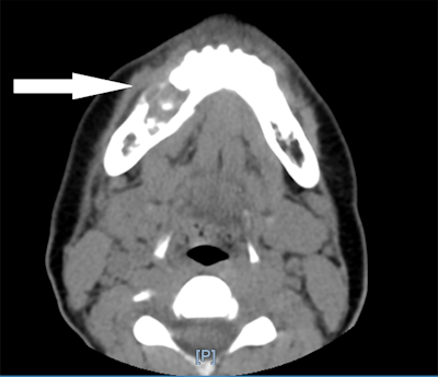 A CT scan without contrast shows a lytic lesion of the right mandible.
Kahlon and colleagues described the case of a 17-year-old who went to a pediatric emergency department after experiencing three days of worsening right jaw pain. He estimated his pain intensity at a nine out of 10 and described it as a dull ache that worsened during brushing or chewing. He experienced some relief after taking ibuprofen, and he reported no other symptoms. Three weeks prior to this visit, his lower right premolar was pulled; he was prescribed antibiotics and sent home.
The lower right first premolar was confirmed missing during examination of the oral cavity. However, there was a 1 x 1-cm tender indurated bonelike buccal vestibular swelling on the lower right jaw adjacent to the missing tooth site. Also, a 5 x 5-mm nonfluctuant pink gum overgrowth lesion was seen at the lower right first premolar site, according to the authors.
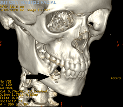 A 3D reconstruction of the scan shows the lytic lesion of the right mandible.
An oral surgery team was consulted, and a CT scan showed a lesion centered in the patient's right jaw with about 2.2 x 1.7 x 1.7 cm of erosion that went through the inner and outer cortex of the mandible. The erosion was centered on the roots of the right lower premolar teeth and first molar tooth with destruction of the second premolar tooth. This suggested a diagnosis of giant cell granuloma, and lab results from a biopsy sample confirmed it.
The surgical team extracted the patient's lower second premolar and removed the lesion with debridement of the mandible, followed by a bone graft. He was given antibiotics and recovered without complications, the authors wrote.


_20210915221258.png?auto=format%2Ccompress&fit=crop&h=167&q=70&w=250)


