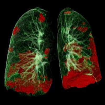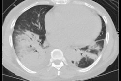
Virtual reality (VR) software can be used to quantify lung damage on the CT exams of COVID-19 patients, potentially helping to guide patient management, according to research presented at this week's virtual Chest 2021 meeting.
Researchers from George Washington University utilized VR software to retrospectively evaluate the CT exams of 27 patients who had been admitted at their institution for COVID-19 over a three-month period last year. They found that those who died had significantly higher volume of infected lung tissue than those who survived.
"Our findings suggest virtual reality is a promising tool for high-resolution evaluation and volumetric analysis of lung injury caused by the 2019 novel coronavirus," said presenter Dr. Keith Mortman, chief of thoracic surgery at George Washington University Hospital.
In their single-center study, the researchers sought to determine the correlation between VR-based quantitative lung damage assessments with patient outcomes -- either discharge or death. They also assessed the correlation of laboratory values at admission with these outcomes in the study, which included 27 consecutive patients admitted with SARS-CoV-2 between March to May 2020 and who had received chest CT scans.
All five lobes of the lung were involved in the majority of these patients. Of the 27 patients, 19 were successfully discharged from the hospital
CT scans were performed on these patients at a median of three days from the onset of symptoms. After generating 360-degree VR scans from each CT exam using VR visualization software (Surgical Theater), the researchers then performed volumetric analysis to calculate lung volume data.
The percentage of diseased tissue was determined by comparing the disease volume with the overall lung volume, normalized for body surface area. The authors were blinded to both clinical and laboratory data during image segmentation.
These results -- along with laboratory data at patient admission -- were then compared with the patient outcomes.
Although there wasn't a significant correlation between the extent of lung infection and outcomes, there was a statistically significant difference in the volume of infected lung tissue between those who survived and those who didn't. This significant difference was also evident when the infected lung volume was normalized to the patient's body surface area.
| Impact of infected lung volume on outcomes of COVID-19 patients | ||
| Patients who were discharged | Patients who died | |
| Mean/median | Mean/median | |
| Infected lung volume (cm3) | 346.32/257.58 | 597.72/628.55 |
| Infected lung volume/body surface area (cm3/m2) | 172.26/106.85 | 304.32/379.67 |
In other findings, the researchers observed significantly elevated blood urea nitrogen (BUN), creatine, and ferritin in patients with a greater percentage of infected lung tissue and the percentage of infected lung tissue normalized to body surface area. In addition, patients with significantly elevated levels of ferritin, glucose, D-dimer, and Interleukin-6 (IL-6), and prothrombin international normalized ratio (INR) were more likely to succumb to the virus.
"To our knowledge, this is the first report of utilization of virtual reality for depicting and quantifying lung injury caused by SARS-CoV-2," Mortman said. "Virtual reality enabled us to better visualize the infected lungs using an immersive 360-degree model."
The researchers also noted that this type of VR analysis could be utilized to establish postinfection baseline results and guide further management of COVID-19 patients.


