Photon-counting CT combined with a deep learning algorithm to correct for noise improves visualization of bone disease caused by multiple myeloma compared with conventional CT, a study published September 6 in Radiology has found.
The results show promise for reducing the radiation exposure to which patients with multiple myeloma may experience, since they often undergo many CT exams over the course of diagnosis, tracking, and treatment, wrote a team led by Dr. Francis Baffour of the Mayo Clinic in Rochester, MN.
"In whole-body low-dose CT, the primary clinical need is to improve the evaluation of skeletal findings while maintaining acceptably low radiation dose to mitigate concerns about radiation risk from repeated scans," the group noted. "At these low doses, [photon-counting] CT is more likely to improve visualization of features of myeloma compared with [conventional] CT."
Up to 80% of patients with multiple myeloma develop bone disease, the researchers explained. Low-dose CT is a sensitive tool for detecting bone lesions, and once bone disease is diagnosed, patients are tracked with further CT imaging to assess progression and guide treatment.
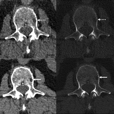
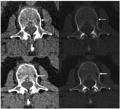
 Reference protocol (top) and evaluated protocol (bottom) images in a 74-year-old man with multiple myeloma. Soft-tissue reconstruction is shown (left side; window width, 400; window level, 40), whereas the right column is the bone reconstruction (right side; window width, 3700; window level, 600). A lytic bone lesion in the L3 vertebral body is more conspicuous on the noncontrast-enhanced axial photon-counting CT reconstruction images (bottom; solid arrows) compared with the noncontrast-enhanced axial energy-integrating detector CT reconstruction images (top; dashed arrows). Image and caption courtesy of the RSNA.
Whole-body low-dose CT has a mean effective dose that ranges between 4 mSv and 8 mSv, but low-dose images can be noisy. Photon-counting CT shows promise for imaging bone disease caused by multiple myeloma because its smaller detector pixel sizes eliminate the need for the high-spatial-resolution filters that conventional CT requires -- which translates not only to better dose efficiency but also less noise.
Baffour and colleagues sought to compare the performance of conventional CT and photon-counting CT for visualizing bone lesions in multiple myeloma patients through a study that included 27 participants who had a conventional CT exam then a photon-counting CT exam between April and July 2021. The photon-counting exam was performed under an ultrahigh-resolution protocol at a radiation dose matched to the conventional CT exam (8 mSv).
Both the conventional and the photon-counting CT images were reconstructed to 2-mm section thickness. The photon-counting images were also reconstructed at 0.6-mm thickness and then reconstructed with a convolutional neural network algorithm that reduced noise.
The investigators found that photon-counting CT had 23% lower image noise compared with conventional CT imaging at the same radiation dose. Assessment of the 2-mm images showed that photon-counting CT enabled visualization of particular bone disease features such as lytic lesions, intramedullary lesions, fatty metamorphosis, and pathologic fractures better than did conventional CT (p = 0.05); the 0.6-mm photon-counting CT images with neural network denoising produced these same results and enabled visualization of more lytic lesions compared with conventional CT in 21 of 27 patients.
The study results could indicate that photon-counting CT could truly improve the care and treatment of patients with multiple myeloma-related bone disease, according to Baffour.
"Our excitement as scientists and radiologists in these results stems from our realization that this scanner could make a difference in the staging of disease, potentially impact therapy choice, and ultimately, patient outcomes," he said in a statement released by the RSNA.


