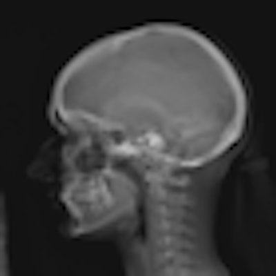
The campaign to reduce pediatric radiation dose has been focusing on CT of late, but efforts are also under way to reduce dose in radiography exams. A study presented this week by Canadian researchers found that a new type of slot-scanning digital radiography (DR) system can produce sharply lower dose in pediatric scoliosis studies compared to computed radiography (CR).
 |
| EOS can scan the entire body in a single study. |
That theory was put to the test in a study of 50 pediatric patients referred for spine radiography to diagnose suspected scoliosis. The study, led by Sylvain Deschênes, Ph.D., of Sainte-Justine Mother and Child University Hospital Center in Montreal, was presented at this week's Journées Françaises de Radiologie (JFR) meeting in Paris.
The researchers used the EOS system on 39 female and 11 male patients with an average age of 14.8 years (± 3.6 years). Patients first received an EOS scan, followed by a CR study (FCR7501S, Fujifilm Medical Systems, Tokyo). In addition to measuring radiation dose, the researchers also assessed the image quality of the EOS system relative to CR.
The study protocol consisted of posterior-anterior and sagittal views of the spine, and images were taken so at least the last cervical vertebra and pelvis were visible. Dosimeters were placed on the skin of each patient to record x-ray dose, and dose for both the EOS system and the CR unit were recorded at seven different locations, ranging from the nape of the neck to the distal iliac crest. Radiation dose was as follows:
Radiation dose, EOS versus CR
|
The researchers commented that entrance dose decreased from a six- to nine-fold reduction with EOS in the thoraco-abdominal region to three times less radiation with EOS at the nape of the neck. They attributed this to the different geometries of the systems: CR produces conic projections, while EOS scans produce cylindrical projections. X-rays travel a shorter distance to reach the neck with EOS, while CR is centered near the thoraco-lumbar junction.
Image quality was rated by four blinded reviewers: two were orthopedic physicians and two were radiologists. They used a four-point scale to rate image quality, ranging from "structure not detectable" to "features clearly defined." Half of the scores rated the systems as having equivalent image quality; where a preference was indicated, most of the scores favored the EOS unit.
Image quality, EOS versus CR
|
Deschênes and colleagues concluded that the EOS system produced better image quality with the benefit of lower radiation dose. One drawback, however, is that the system's slot-scanning architecture produces a uniform radiation dose across the entire area of the body being imaged, even though the patient's body thickness varies.
The researchers concluded by stating that for their next study they intend to compare EOS to a digital radiography system -- initial findings indicate that EOS images are as good or better 83% of the time, they said. They also intend to study the use of the system for following up lung nodules.
By Brian Casey
AuntMinnie.com staff writer
November 3, 2008
Related Reading
French firm Biospace Med prepares for launch of EOS digital x-ray unit, December 25, 2006
Copyright © 2008 AuntMinnie.com


