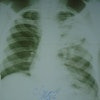
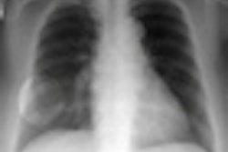
All hospitals aim to keep radiation dose exposure to a minimum when taking chest radiographs of infants, but this goal can be undermined by invisible "dose creep" of digital x-ray equipment. An article recently published in Pediatric Radiology describes an easy way to monitor exposure levels.
Pediatric radiologists at the Riley Hospital for Children in Indianapolis are using an exposure index and deviation index developed by the International Electrotechnical Commission (IEC) in Geneva and the American Association of Physicists in Medicine (AAPM) in College Park, MD, to monitor exposure drift of the radiography systems they use to acquire neonatal chest radiographs (Pediatric Radiology, December 30, 2010).
Although the use of portable computed radiography (CR) and digital radiography (DR) equipment has dramatically increased the efficiency of taking chest x-ray exams of premature and newborn infants in neonatal intensive care units and nurseries, it has made monitoring dose exposure more difficult. With x-ray film, it's easy to identify overexposed images. But because of the wide dynamic range of digital x-ray systems, the postprocessing of overexposed images generally results in no appreciable difference that can be detected by the eye.
Exposure indexes for each examination type and body part are now incorporated into CR equipment to show radiation dose exposure levels and are displayed on individual images. Target exposure indexes can vary by examination room, as they are dependent upon factors such as filtration and sensitivity of the detector plate. For this reason, the actual value of the exposure index cannot be used to track patient dose.
However, a deviation index can be used to calculate the difference between a desired target exposure and the actual exposure, and this value can be displayed automatically in a PACS for each x-ray image. This enables data to be collected and analyzed by patient and by exam type.
|
Calculation of the deviation index: Deviation index = 10 x [Log10 (exposure index/target exposure index)] |
Mervyn Cohen, MD, a pediatric radiologist and professor of radiology at the Indiana University School of Medicine, and colleagues conducted a study to evaluate whether the exposure and deviation indexes could be used for routine quality assurance monitoring.
Using the department's existing exposure method, which optimized radiation dose for neonatal chest exams, the researchers created a target exposure index as the mean of 50 chest images for infants of three sizes. They then calculated the exposure/deviation indexes for 1,884 consecutive images acquired over a 90-day period. A phantom simulating the chest of a 1,500-g infant was imaged on a weekly basis as a control.
Ninety-three percent of the exposures were within three deviation units. The calculated increase in mean weekly exposure was less than 10% for patient images and 8% for the phantom.
Expect variation in the exposure index, the authors commented: Infants vary in size, tube output may differ, and anode-detector distances may vary due to problems in positioning the portable x-ray machine around different incubators in the neonatal nurseries. Because variation by individual technologist can also be monitored, technologist-specific errors can be rapidly and easily identified.
Based on the study results, the authors concluded that exposure and deviation indexes offer an excellent method of tracking radiation exposure from CR systems for infants and similarly sized children, for a specific body part, for a specific imaging system, and when the kVp is kept relatively constant. They have incorporated use of the index into the department's quality assurance program.
By Cynthia E. Keen
AuntMinnie.com staff writer
January 27, 2011
Related Reading
Exposure index varies widely between CR systems, April 9, 2010
X-ray scatter radiation risk is low to NICU infants, January 12, 2010
QA application can bolster DR image quality, consistency, December 22, 2008
Copyright © 2011 AuntMinnie.com


