By allowing radiologists to more easily spot lesions that overlap with bone, a bone suppression software application yielded a significant increase in the detection of lung nodules on digital x-ray exams, according to research published online April 14 in Radiology.
In the study involving 15 radiologists and 368 patients, a team led by Dr. Matthew Freedman of Georgetown University found that the software increased reader sensitivity by nearly 17%, albeit with a slight but significant decrease in specificity. Most of the newly detected lesions had a high degree of overlap by bone.
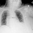
"Chest radiographs, after software processing, can now be viewed with the ribs and clavicles less visible and with the lung structures contrast-equalized," the authors wrote. "Patients are more likely to have lung nodules detected when software is used."
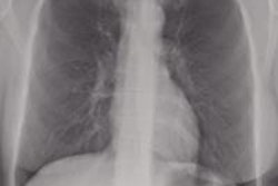
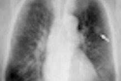
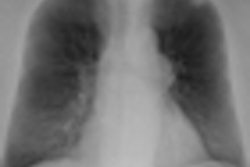
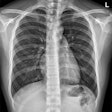
The researchers evaluated SoftView software (Riverain Medical), which is designed to aid radiologists in detecting lung nodules and other lung cancer findings. It produces a modified image by suppressing the ribs and clavicles, and it also filters noise and equalizes contrast in the area of the lungs, according to the authors.
Fifteen radiologists with a postresidency experience range of 0.1 to 27 years were recruited to participate in the retrospective study. Of the 368 cases, 266 were digitized film-screen studies, 66 were computed radiography (CR) exams, and 19 were digital radiography (DR) studies. There were 122 patients with proven lung cancer, and 246 confirmed to be cancer-free.
The radiologists were presented with an individually randomized sequence of images, with each case consisting of a standard frontal chest radiograph and a software-enhanced image.
The radiologists then marked the location of the most suspicious nodule, if any. After interpretation was completed on the standard radiograph, they viewed the software-enhanced image and again marked the location of the most suspicious nodule, if any. For both images, the radiologists also provided the level of suspicion on a scale of 0 (nonactionable) to 100 (absolutely actionable).
They also chose to recommend or not recommend further investigation -- with either CT or biopsy -- based on the standard image alone and then based on the software-enhanced image. These data were used to determine sensitivity and specificity for nodule detection, according to the authors.
For all readers, the mean area under the localized receiver operator characteristics (LROC) curve was 0.460 (95% confidence interval [CI]: 0.435-0.486) for conventional images and 0.558 (95% CI: 0.539-0.578) for software-enhanced images. The mean change was statistically significant (p = 0.0001).
The authors also discovered a nearly 17% increase in sensitivity after using the software-enhanced images (p < 0.0001), but there was a significant decline in specificity (p = 0.004).
Bone-suppression software versus standard images
|
In other findings, approximately 65% of the new detections with the software were in those with 80% or greater bone overlap, and approximately 74% were in those with 70% or greater bone overlap, according to the authors.
| All radiologists participating in the study received a set payment from Riverain, but payment was not based on their results. Georgetown University reported receiving payments from Riverain for writing and reviewing the current manuscript, and the university also reported receiving equipment and software on loan from Riverain. |


