Increasing the source-to-image distance (SID) for digital radiography (DR) examinations of the pelvis reduces radiation dose while maintaining image quality, according to a study published in the September/October 2011 issue of Radiologic Technology, a journal of the American Society of Radiologic Technologists (ASRT).
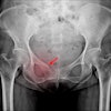
Andrew England from the University of Liverpool and colleagues investigated distance, dose, and image quality for DR exams of the pelvis, the second most common bucky procedure in the country.
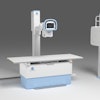
To perform the study, England's team positioned an anthropomorphic pelvic phantom for a standard anteroposterior examination. Using a single DR unit with a flat-panel digital detector, the group set an initial source-to-image distance of 100 cm and used an antiscatter focused radiation grid. The x-ray beam was collimated for a standard anteroposterior pelvic examination and was kept constant throughout the experiment, while source-to-image distance varied from 80 cm to 147 cm.
The results indicate that by increasing the SID, both the entrance surface dose and effective dose decreased when using the antiscatter radiation grid, and further decreased when the grid was removed. At a 147-cm source-to-image distance, the decrease in entrance surface dose and effective dose when using a grid equated to a 25% reduction in radiation dose compared to standard parameters.
