Japanese researchers have found that combining two advanced digital radiography (DR) techniques -- tomosynthesis and energy subtraction -- results in major improvements over conventional digital x-ray energy subtraction when imaging pulmonary nodules, according to an article in the November issue of Academic Radiology.
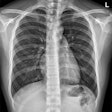
Traditional x-ray has never performed well as a tool for finding pulmonary nodules, due to the fact that it's a 2D representation of a 3D volume, where structures such as rib bones can overlap and superimpose on lung tissue, according to a research team led by Tsutomu Gomi, PhD, of Kitasato University. CT has developed into the gold standard for nodule detection, but it comes with a higher cost, slower workflow, and a higher radiation burden, they wrote (Acad Radiol, Vol. 20:11, pp. 1357-1363).
New advanced digital x-ray techniques hold promise that radiography could improve its performance in the chest. And who knows? Perhaps someday DR could become a screening tool to rival CT for pulmonary nodule detection.
Advanced x-ray options
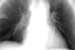
One of the advanced x-ray techniques is dual-energy subtraction (DES), which was first pioneered years ago in computed radiography (CR) as a technique to compensate for the superimposition issue. With energy subtraction, images are acquired at two separate energy levels; because different types of tissue respond to x-ray energies in different ways, the images can be digitally "subtracted" to produce a single image that's free of interference from bones.
In recent years, digital tomosynthesis has emerged as another alternative in DR for dealing with superimposed structures. With the technique, tomographic images are acquired in an arc around the patient, and then reconstructed into a volume that enables radiologists to see around structures that might be obscuring pathology.
Gomi and colleagues posited that combining energy subtraction with tomosynthesis could produce a technique that's even more powerful than either tool working alone. After previous research on phantoms, they decided to test their theory by performing dual-energy subtraction on a tomosynthesis DR unit (DES-DT) and comparing it to dual-energy subtraction on a conventional DR system (DES-R) in a group of 36 patients.
The study group was drawn from patients with pulmonary nodules who had been referred for chest CT. There were 19 men with a mean age of 69 years in the group, and 17 women with a mean age of 66 years.
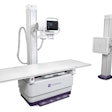
For the dual-energy tomosynthesis studies, imaging was performed on a commercially available unit (SonialVision Safire II, Shimadzu Medical Systems). Energy levels of 60 kVp and 120 kVp were used for the dual-energy part of the acquisition, and the system had a scan time of 6.4 seconds and a swing angle of 40°. Radiation dose was approximately 1.22 mSv.
For the DES-R portion of the study, dual-energy images were acquired on a commercially available DR system (CXDI-50C, Canon) at 60 kVp and 120 kVp, with an effective radiation dose of about 0.39 mSv. Images from a 64-slice CT scanner served as the reference standard; radiologists noted lesion location and measured lesion size on the CT scans, classifying them according to Fleischner criteria.
Both DES-DT and DES-R images were interpreted by five readers, consisting of two radiologists and three doctors of pulmonary medicine. In addition to the 36 cases with nodules, the researchers created 36 cases without lesions.
The readers were asked to assess the DES-DT and DES-R images and rate their confidence that each case contained a pulmonary nodule. The researchers then used receiver operator characteristics (ROC) analysis to assess the performance of the readers by criteria such as sensitivity, specificity, accuracy, and lesion detectability (as measured by area under the curve, or AUC).
Gomi and colleagues found that dual-energy subtraction with the tomosynthesis technique produced images with greater contrast than dual-energy radiography.
"When the nodules were no longer superimposed over the normal structures, their characteristics and distribution could be observed much more clearly," they wrote.
Relevant statistical analysis for the technique as averaged across all five readers showed superiority for DES-DT in all categories except specificity, as indicated below.
| DES-DT vs. DES-R for pulmonary nodules | ||
| DES-DT | DES-R | |
| Sensitivity | 87.7% | 53.8% |
| Specificity | 78.3% | 78.4% |
| Accuracy | 83.1% | 66.1% |
| Lesion detectability (AUC) | 0.94 | 0.76 |
CT found all 36 nodules, of which 80% were found on DES-DT and 58% were found on DES-R. In all, 32 of the 36 nodules were larger than 8 mm, two were 6 to 8 mm, and two were 4 to 6 mm. No nodules smaller than 4 mm were found.
In discussing their results, the researchers pointed out the novelty of dual-energy subtraction tomosynthesis, and to their knowledge, there is "no history of evidence" supporting its integration into clinical practice beyond the current study. They concluded by stating that DES-DT was able to confirm true pulmonary lesions and was a clear improvement over dual-energy radiography, which demonstrated low sensitivity and a large number of false-negative findings compared to CT.
They recommended future research with larger patient populations, as well as studies of dual-energy tomosynthesis in patients with lesions smaller than 8 mm.


