Thursday, December 5 | 12:15 p.m.-12:45 p.m. | CL-PDS-TH2A | Room S101AB
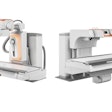
In this poster presentation, researchers will discuss how an ultralow-dose protocol using a biplanar slot-scanning digital x-ray system with optimized acquisition parameters can be used to monitor idiopathic scoliosis, allowing further dose reduction.Biplanar slot scanning has been shown to significantly reduce dose when monitoring spinal deformities. In addition, dose can be reduced even further with recent technical advances such as copper filtration and dedicated image processing, as well as with the optimization of acquisition parameters (kV, mA, and scan speed).
In this study, 23 patients (mean age, 12.4 years) with mild or moderate idiopathic scoliosis were imaged with an ultralow-dose protocol optimized according to body mass index. Dose area product (DAP) and entrance dose (kerma) were quantified for each examination. Image quality was rated on a five-point scale based on the visibility of the edges of the vertebrae in five different anatomical areas.
Mean DAP was 39.5 (± 17.1) mGy-cm2 and 87.9 (± 31.7) mGy-cm2 for the anteroposterior (AP) and lateral views, respectively. Mean entrance dose was 17.6 (± 6.4) µGy and 42.1 (± 12.8) µGy for the AP and the lateral views, which corresponds to approximately 10 days of background radiation, according to Dr. Guy Sebag from Robert-Debré Hospital in Paris. Image quality was graded 3 in 48% of cases, 4 in 48% of cases, and 5 in the remaining 4% of cases.
The study findings prove that ultralow-dose imaging is achievable for the follow-up of idiopathic scoliosis, with acceptable image quality and high reproducibility of the measurements, according to the researchers.
Sebag and colleagues plan to assess the reproducibility of scoliosis parameter measurement with 3D reconstruction, he said in an interview with AuntMinnie.com.


