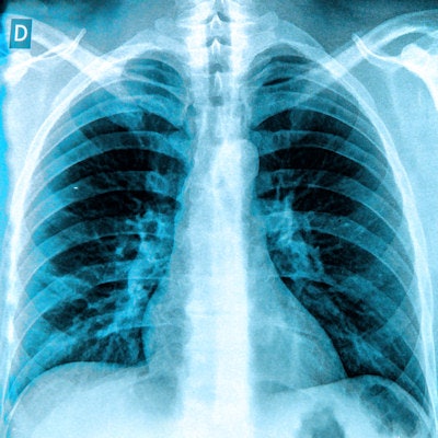
Researchers in Japan have refined an algorithm that someday could be used to reduce patient recognition and identification errors in digital x-ray studies. They tested their enhanced algorithm and found that it scored 100% for recognizing the correct orientation of digital x-ray images.
The group from Kyushu University, led by Yoichiro Shimizu, tested an algorithm that automatically extracts and analyzes what the authors call "biological fingerprints" in images, consisting of different image characteristics. They used the fingerprints to create an "averaged" digital chest that could serve as a ground truth image to ensure that images acquired in clinical settings are correctly oriented.
Shimizu and colleagues believe the algorithm ultimately could be used to automatically detect and correct image orientation errors, which occur with wireless digital radiography (DR) and with larger DR panels. Their results were published April 27 in Radiological Physics and Technology.
Biological fingerprints
In a previous paper published in 2013, the researchers described their development of an algorithm designed to help with quality control in digital x-ray, such as by finding lost x-ray exams in a PACS archive. The technique matches images by analyzing five different characteristics, such as cardiac shadow and right lower lung attributes; in the 2013 study, the technique was used to find misfiled images and match them with correct images.
In the current study, the group applied the algorithm to another digital x-ray headache: images that are misoriented. This can occur if a wireless flat-panel detector is not used in the correct orientation -- for example, in cases of bedside radiography.
To test the algorithm, the researchers pulled 200 test images (100 men and 100 women) at random from a database of more than 36,000 chest radiographs acquired with computed radiography as part of a lung cancer screening program in the Iwate prefecture. They used these to create what they called averaged images -- images that represented the average thoracic anatomy based on a combination of features across all the images. An average image for each gender was created based on the 100 images of each.
They then acquired another 200 images from the database to create target images, the images that would test the accuracy of the algorithm. Finally, they compared the target images with the average images, using the positions of the five biological fingerprints to determine whether the target images were oriented correctly.
They found that the algorithm was able to automatically and precisely extract biological fingerprints in all 200 cases (100%). They also found that the enhanced version of the algorithm was superior to previous versions in correctly extracting the five different fingerprints.
The group believes that the algorithm could also be useful for analyzing images acquired with different sizes of flat-panel detectors. Some technologists using larger detectors pay less attention to patient positioning than with a smaller detector; use of the biological fingerprint algorithm could compensate for this.
"We believe that the proposed method of utilizing an averaged image for each sex is useful for recognizing image rotation of chest radiographs and for extraction of [biological fingerprints], especially from chest radiographs acquired by larger x-ray detectors," they wrote. "Our method could also be applied to bedside chest radiographs."
Shimizu and colleagues noted that one limitation of the study is the need to know the sex of the patient before the algorithm can be applied, since the averaged images differ by gender. In a preliminary experiment, they found lower accuracy for the algorithm in trying to match images of different genders, most likely caused by breast shadow in women.
Also, there can be differences in mediastinum and cardiac shadows between anteroposterior (AP) and posteroanterior (PA) images, with AP images having wider shadows. This would have to be addressed in future iterations of the algorithm, they wrote.


