 Chris Schmidt.
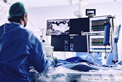
Cardiac interventional (CI) radiographers play a vital role in the diagnosis and treatment of patients during their procedure in the cardiac catheterization laboratory. They are skilled healthcare professionals who specialize in using imaging technology and minimally invasive procedures to diagnose and treat various cardiac and vascular conditions.
CI radiographers work closely with radiologists, vascular surgeons, and interventional cardiologists to perform procedures such as angioplasty, stent placement, heart perfusion studies, and more. These procedures are often used to open blocked blood vessels, repair damaged vessels, or treat other cardiovascular issues.
CI procedures are often done with an endovascular approach, which means they access within the vessel. This is done using the Seldinger Technique where a wire is introduced into an artery or vein allowing the physician to locate the anatomy of choice by navigating through the vessels. This is a minimally invasive procedure that leaves a two-millimeter incision on the patient's skin that will quickly heal over with minor restrictions.
Prior to the development of endovascular treatment, patients who needed stenting or vessel repair would need complex open surgeries that could have multiple complications with long hospital stays and a long road to recovery. Now, many cardiac interventions are done as outpatient procedures that require no overnight stay and have minimal recovery time.
Improving patient outcomes
A competent CI radiographer can improve the success rate of exams and improve patient outcomes. The overall success of a procedure has a lot to do with the skill of the CI radiographer. CI radiographers will go through extensive training to learn cardiac procedures and to be able to assist physicians. They are trained in advanced cardiac life support, EKG interpretation, coronary anatomy, vascular anatomy, and hemodynamics.
A physician will expect a CI radiographer to notify them if there is a sudden heart rhythm change, a drop in blood pressure, or a waveform change that could indicate a damping of the catheter inside the vessel. The damping of a waveform could indicate a fully obstructed vessel caused by the catheter restricting blood flow.
If this goes unnoticed, the patient may develop a fatal rhythm or their vitals might desaturate. The more in tune a CI radiographer is to a procedure, the better patient care they can offer by preventing these rhythms from becoming fatal and making sure the hemodynamics are stable for proper perfusion.
Helping with diagnosis
A CI radiographer also helps with the diagnosis of a patient. During a potential coronary intervention, the physician will have the CI radiographer operate the diagnosing equipment. Physicians are often unfamiliar with how to proficiently run these machines, so they rely on the capability of the CI radiographer.
Intravascular ultrasound and optical coherence tomography are two examples of these diagnostic tools. These tools accurately measure vessel size, the percentage of occlusion, and look for acoustic shadowing that could indicate calcification that would change the course of intervention. How well the CI radiographer knows how to operate this equipment impacts information that may determine treatment, including the correct balloon size, stent size, and if the stenosis is treatable or if the patient will need open heart surgery.
CI radiographers must have precision and accuracy when intervening with the physician. Every balloon must be prepped before use, catheters must be flushed, and the CI radiographer must know what anatomy they are looking at while injecting contrast into the vessel. If the balloons and catheters are not properly flushed or prepped, there is a risk of sending an air embolism to the patient.
CI radiographers must also be aware of the vessel size and the engagement of the catheter into the vessel. They are responsible for injecting contrast into the vessel to optimize visualization and must use the correct rate of injection and pressure so the physician can properly inspect the entire vessel.
If they do not get enough contrast, there is a risk of a streaming artifact that could miss a stenosis in a coronary artery. If the CI radiographer injects too forcefully, they risk losing engagement with the vessel. The physician would then need to re-cannulate the artery, which can increase the procedure time and reduce the chance of success.
Stent deployment
An important task when it comes to CI treatment is the deployment of a stent. The physician lines up the stent in the correct location, and to prevent catheter, wire, or stent movement, holds these items together. The CI radiographer then must know how to properly deploy the stent, reach optimal inflation, and be prepared to re-inflate in a second’s notice. This is one of the most dangerous parts of a percutaneous coronary intervention. There is always a minimal risk of coronary artery perforation during a balloon and stent deployment.
The sterile table must be organized at all times and ready to respond with multiple different stents and balloons if needed. CI radiographers are proficient at multitasking, as they must always be prepared to respond to an emergency during the procedure, continue to monitor vitals, perform contrast injections, always be aware of the total contrast amount, and keep the table organized to assist in many ways.
The CI radiographer constantly practices radiation safety by collimating the x-ray exposure, reducing the object-image distance (OID) by minimizing the distance from the flat panel detector to the patient, thus improving the spatial resolution of the image.
Patient compassion
CI radiographers also display patient compassion. They are typically one of the first ones to interact with the patient when they come into the laboratory and they are usually one of the last people the patient will see, as the CI radiographer will either pull the arterial sheath from a heart catheterization or will give the patient proper instruction that will reduce possible bleeding.
In summary, CI radiographers are integral members of the healthcare team and play a vital role in diagnosing and treating heart and vascular diseases with precision, compassion, and a commitment to patient safety.
Their expertise in minimally invasive procedures is instrumental in improving cardiac care and enhancing patient outcomes. Their extensive training ensures that CI radiographers can perform their important work to the highest level of competency and safety
Chris Schmidt is program director at Baker College of Muskegon and a cardiac interventional technologist.
The comments and observations expressed are those of the author and do not necessarily reflect the opinions of AuntMinnie.com.


