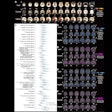
An international team of psychiatrists used PET imaging to examine the underlying cause of tics in patients with Tourette syndrome (TS). While tics are the defining symptom of TS, the neurological mechanics behind them are poorly understood, according to the group’s paper in the August issue of the Archives of General Psychiatry.
"To date, the functional neuroimaging experiments of TS have provided extremely valuable information, but have not … generated an image of the brain state specifically associated with tics," the authors wrote (Archives of General Psychiatry, August 2000, Vol.57, pp.741-748).
The group consisted of clinicians from Weill Medical College of Cornell University in New York City, the Institute of Neurology and National Hospital for Neurologic Diseases in London, and the Prince Henry Hospital in Sydney.
Six male patients with a diagnosis of TS made up the study population. Two patients were not on medication, while four suffered from tics despite neuroleptic medication.
Regional cerebral blood flow was measured with a Siemens 953B PET scanner (Siemens Medical Systems, Hoffman Estates, IL). The scans were performed in high-sensitivity 3-D mode with a low-dose, [5O]H2O slow-bolus technique. The acquisition time was 90 seconds, including a critical 30 seconds during which the pattern of radiotracer distribution in the brain was determined.
According to the results, the scans showed extensive activity in areas of the brain generally associated with the selection, preparation, and initiation of behavior, the group stated.
"Increased brain activity highly correlated with tic behavior was detected in a set of neocortical, paralimbic, and subcortical regions," they determined, including several areas of the primary motor cortices and the Broca’s area.
"The extensive activity in executive and premotor regions may be particularly notable, and may help to extend our understanding of disordered action and volition in TS," they wrote. More specifically, the tics may represent a paradoxical state in the brain where areas that are normally associated with a subjective sense of volition are not operating under the volitional control of the patient, they wrote.
Three sets of circuits involved in the selection and initiation of movement also were pinpointed as areas of high activity: the motor, dorsolateral prefrontal, and anterior cingulate circuits. Modulation of these circuits with dopamine blockers or other treatment could aid in controlling tics, they said.
By Shalmali PalAuntMinnie.com staff writer
August 30, 2000
Let AuntMinnie.com know what you think about this story.
Copyright © 2000 AuntMinnie.com


