Japanese researchers are using PET to visualize copper distribution in mice to potentially identify therapies for a genetic disorder known as Menkes disease.
The study, led by Dr. Shiho Nomura of Osaka City University and published online March 13 in the Journal of Nuclear Medicine, could lay the groundwork for PET studies of human Menkes patients to identify new therapy options.
Menkes is a rare disorder in which individuals are unable to absorb copper; most affected babies die within the first few years of life. The current standard treatment is to inject copper, but the therapy has limited efficacy and eventually becomes ineffective. This leads to neurodegeneration, and copper accumulates in the kidneys, sometimes leading to renal failure, according to a release from Riken Center for Life Science Technologies, with which several co-authors are affiliated.
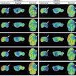
In the study, the researchers injected the radioisotope copper-64 into Menkes disease model mice, which have an inborn defect in copper metabolism. They then used PET to visualize how the copper moved throughout the body during a four-hour period. They compared mice injected with copper alone to mice injected with copper along with one of two drugs: disulfiram or D-penicillamine.
The mice given copper and disulfiram had a relatively high concentration of copper in the brain without a significant increase in the kidneys. In addition, the amount of copper going to the brain in mice treated with disulfiram was greater than in those treated with copper alone, suggesting that the drug has an effect on the passage of copper through the blood-brain barrier, according to the researchers.


