Tuesday, November 27 | 3:50 p.m.-4:00 p.m. | SSJ03-06 | Room E353A
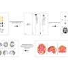
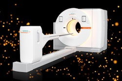
Simultaneous cardiac FDG-PET/MRI could be a welcome option for the early detection of cardiac involvement in Anderson-Fabry disease and identifying different stages of disease progression, according to researchers from Italy.Dr. Andrea Ponsiglione from University Federico II in Naples, will present findings from 17 women with Anderson-Fabry disease and normal left ventricular (LV) function. The patients underwent cardiac PET/MRI (Biograph mMR, Siemens Healthineers) after administration of FDG and gadolinium-diethylenetriamine penta-acetic acid (DPTA) for assessment of late gadolinium enhancement assessment, which is an indication of intramyocardial fibrosis. Five patients (29%) showed focal late gadolinium enhancement and were excluded from the final analysis.
Mean MRI T1 values were significantly lower in patients with Anderson-Fabry disease than in control subjects. With PET, seven patients (58%) showed abnormal coefficient of variation values, which suggest a pattern of inflammation. The other five women (42%) showed normal coefficient of variation values with homogeneous FDG uptake. Patients with abnormal coefficient of variation values also showed higher mean T1 values of lateral segments of the mid-LV wall, which also could indicate a correlation between progressive myocyte sphingolipid accumulation and inflammation.
The study shows the potential role of FDG-PET/MRI in the early detection of cardiac involvement in patients with Anderson-Fabry disease and also monitoring disease progression, according to Ponsiglione and colleagues.


