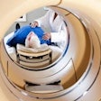
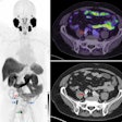
Who says PET is only for humans? Researchers at the University of California, Davis (UC Davis) School of Veterinary Medicine are using a prototype PET scanner to image the hooves of horses in a unique way.
The equine PET device allows a horse to stand upright, rather than on its side, with scans performed while the animal is under sedation rather than anesthesia, according to a January 27 article in Horsetalk (naturally).
With this approach, a clinician places the sedated horse's hoof into an open hole in the horizontal scanner, which sits on the floor and contains a ring of PET detectors. The design is low enough for the horse to step out safely. The scanner creates 3D PET images within five minutes, with the help of sodium fluoride to image bone and FDG for soft-tissue imaging. The results confirmed PET's efficacy for imaging the horse in the standing position with no downgrade in image quality from motion artifacts, according to the researchers.
The device was originally developed by Brain Biosciences of Rockville, MD, to image the human brain. The UC Davis researchers believe that this equine PET application will expand to more routine use in veterinary circles and potentially for human patients who cannot undergo anesthesia.


