Japanese and German researchers are reporting early success with a PET imaging agent that could both diagnose and treat patents with prostate cancer, according to a preclinical study published in the November issue of the Journal of Nuclear Medicine.
The study, led by Dr. Fumihiko Soeda from Osaka University Graduate School of Medicine in Japan, was designed to evaluate the change in the whole-body distribution of the theranostic imaging agent fluorine-18 (F-18) prostate-specific membrane antigen (PSMA)-1007 when solutions with different peptide concentrations are used (JNM, November 2019, Vol. 60:11, pp. 1594-1599.)
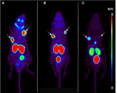
Soeda and colleagues used a mouse xenograft model for three groups of mice, which were divided by the amount of peptide injected into each subject. A decrease in the peptide concentration (or molar activity) level resulted in decreased uptake in the tumor and, to a greater degree, in the salivary glands.
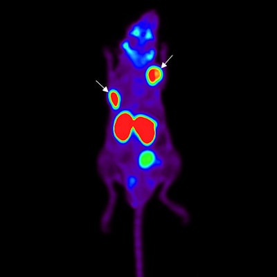
 Maximum intensity projections of F-18 PSMA-1007 PET images in xenografts of mice with high (A), medium (B), and low (C) concentrations of the agent. Tumors are indicated by arrows. Tumor uptake was lower in the low-concentration group, compared with the other two sets of mice. Images courtesy of Soeda et al and the Journal of Nuclear Medicine.
The researchers found that the salivary glands showed a more sensitive decrease in uptake, compared with a reduced molar activity level of the radioligand solution than the tumors.
"This suggests that molar activity is an essential factor to define the optimal distribution for theranostic compounds," Soeda said in a statement released by the Society of Nuclear Medicine and Molecular Imaging. "There is a possibility of obtaining a therapeutic effect in the tumor, while minimizing the adverse effects in the salivary glands by setting an appropriate molar activity level in PSMA-targeted therapy."


