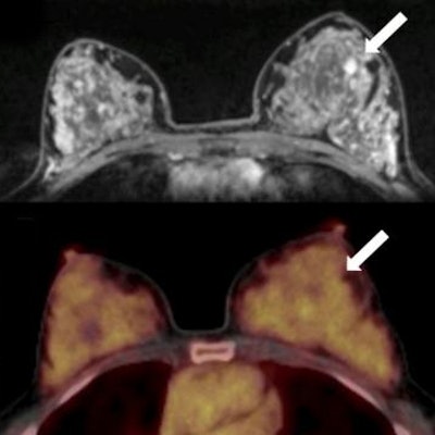
In a finding they described as "unexpected," researchers used PET and MRI to discover that women with a malignant tumor in one breast have lower levels of certain biomarkers in the contralateral, healthy breast. Results were published in the January edition of the Journal of Nuclear Medicine.
The researchers noted that both PET and MRI are increasingly being used in the clinical setting, such as with hybrid PET/MRI scanners, combining the advantages found in both modalities for breast imaging. Biomarkers like background parenchymal enhancement and physiological activity of fibroglandular tissue on MRI and breast parenchymal uptake of FDG on PET could help reveal the presence of malignant tumors -- particularly as these biomarkers occur in healthy breast tissue (JNM, January 2020, Vol. 61:1, pp. 20-25).
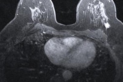
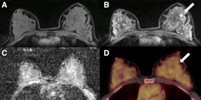 A 50-year-old postmenopausal woman with fibroadenoma (arrows) in left breast. (A) Unenhanced fat-saturated T1-weighted MRI shows extreme amount of fibroglandular tissue (ACR d). (B) Moderate background parenchymal enhancement is seen on dynamic contrast-enhanced MRI at 90 sec. (C) Mean ADC of breast parenchyma of contralateral breast on diffusion-weighted imaging with ADC mapping is 1.5 x 10⁻³ mm2/sec. (D) On F-18 FDG PET/CT, lesion is not F-18 FDG-avid, and background parenchymal uptake of normal breast parenchyma is relatively high, with SUVmax of 3.2.
To that end, a multi-institutional research team examined 141 women seen at the University of Vienna who had imaging abnormalities found on mammography or sonography in one breast and had a tumor-free contralateral breast. Their goal was to determine whether suspicious lesions in one breast could be characterized using biomarkers provided by PET/MRI scans of the contralateral breast. The lead study author was Dr. Doris Leithner of Memorial Sloan Kettering Cancer Center in New York City.
There were 100 malignant and 41 benign lesions in the study population. PET was performed on a PET/CT system (Biograph 64 TruePoint, Siemens Healthineers), while MRI was conducted on a 3-tesla scanner (Magnetom Tim Trio, Siemens). The biomarkers that were assessed and the modality from which they were derived were as follows:
- MRI: Background parenchymal enhancement
- MRI: Fibroglandular tissue
- MRI: Mean apparent diffusion coefficient (ADC) (from diffusion-weighed imaging [DWI] scans)
- PET: Breast parenchymal uptake (from F-18 FDG PET)
The researchers found statistically significant differences in background parenchymal enhancement and background parenchymal uptake in the healthy breast in women with malignant lesions compared with those with benign lesions. Differences in fibroglandular tissue did not achieve statistical significance, nor did differences in ADC mapping on the DWI-MRI scans.
The study authors described the findings as "unexpected," particularly as previous studies have found that breast cancer risk increased in women with higher levels of background parenchymal enhancement. They speculated that previous studies focused on high-risk women, whose breast tissue is known to differ from women of average cancer risk.
If the current findings are confirmed in other studies, the authors believe that there could be relevant clinical applications. For example, women who have a decrease in background parenchymal enhancement and breast parenchymal uptake in the contralateral breast over time might be referred to short-term follow-up to detect early-stage cancer.
"At this point, the underlying processes for the results of our study remain unclear and must be confirmed by studies with larger numbers of individuals at average cancer risk," Leithner and colleagues concluded.


