FDG-PET/MRI is comparable or superior in diagnostic performance to FDG-PET/CT across a range of cancers and endpoints, according to a review of head-to-head studies published July 17 in the American Journal of Roentgenology.
A group led by Amit Singnurkar, MD, of the University of Toronto in Ontario, Canada, reviewed 29 published studies featuring 1,656 total patients with a range of cancer types. They found that FDG-PET/MRI showed comparable or even better performance relative to FDG-PET/CT across a range of diagnostic endpoints.
“The findings help to identify clinical settings where PET/MRI may provide particular clinical benefit for oncologic evaluation,” the group wrote.
Since the last systematic review comparing the methods in 2017, evidence on the use of FDG-PET/MRI using hybrid systems has grown substantially, the authors noted.
To provide the latest evidence, the researchers queried Medline, Embase, and the Cochrane Database of Systematic Reviews (via OVID) for head-to-head comparison studies between the two.
The findings included the following:
- For patient-level detection of regional nodal metastases (5 studies), pooled sensitivity and specificity were 88% and 92% for PET/MRI compared with 86% and 86% for PET/CT.
- For lesion-level detection of recurrence and/or metastases (5 studies), pooled sensitivity and specificity were 94% and 83% for PET/MRI compared with 91% and 81% for PET/CT.
- PET/MRI showed a staging accuracy in breast cancer of 98% versus 74.5% for PET/CT and in colorectal cancer a staging accuracy of 96.2% versus 69.2%.
- PET/MRI sensitivity for primary tumor detection in cervical cancer was 93.2% versus 66.2% for PET/CT.
| PET/MRI vs. PET/CT for lesion-level liver metastasis detection | ||
|---|---|---|
| Measure | PET/CT | PET/MRI |
| Sensitivity | 42.3% to 71.1% | 91.1% to 98% |
| Specificity | 83.3% to 98.6% | 100% |
| Accuracy | 44.7% to 86.7% | 96.5% to 98.2% |
In addition, in three studies, patient management was more commonly impacted by information from PET/MRI (5.2-11.1%) than PET/CT (0.0-2.6%), the authors noted.
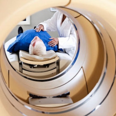
Ultimately, integrated PET/MRI systems combine the advantages of FDG-PET with the advantages of MRI through true temporally and spatially simultaneous acquisitions, the authors wrote. However, despite its benefits, the broad clinical adoption of PET/MRI faces ongoing challenges, they noted.
PET/MRI scanners are more expensive than PET/CT scanners, for instance, and currently have limited clinical availability, with approximately 30 PET/MRI systems versus more than 1,600 PET/CT systems installed in the U.S., according to the authors. Also, examination times are longer for PET/MRI than for PET/CT.
“Standardized PET/MRI protocols are needed to help streamline examinations and limit acquisition times, as well as to promote the quality and consistency of PET/MRI across centers. A multi-society consensus recommendation from 2023 on the use of PET/MRI in oncology should help to achieve this aim,” the group concluded.
The full article is available here.




![Current PET/MRI imaging of healthy and damaged mouse kidneys using the dual contrast agent F-18 [Gd (FL1)]. The imaging provides consistent signals from both PET and MRI. In healthy kidneys, the center of the kidneys appears dark in the MRI image and very bright in the PET image, indicating normal excretion of the contrast agent. The onset of impaired function in one kidney is indicated by the accumulation of the contrast agent and its slow excretion. Precise mapping of MRI and PET data ensures detailed morphological and quantitative analysis of kidney impairment. Image and caption courtesy of IOCB Prague.](https://img.auntminnie.com/files/base/smg/all/image/2024/08/PET_MRI_agent.66b25358df3b3.png?auto=format%2Ccompress&fit=crop&h=167&q=70&w=250)


