Hybrid PET/MRI scans outperform either technique alone for identifying tumors in high-risk patients with suspected prostate cancer, according to research presented February 27 at ECR in Vienna.
The finding is from a preliminary analysis of 23 patients enrolled in an ongoing clinical trial, noted Giorgio Brembilla, MD, PhD, of the IRCCS San Raffaele Scientific Institute in Milan, Italy.
 Giorgio Brembilla, MD, PhD.
“Our preliminary data confirmed the potential increase of sensitivity of the combined use of PSMA PET with MRI compared to each modality alone. We observed 100% negative predictive value,” he said.
Multiparametric MRI (mpMRI) is a widely used approach for diagnosing patients with prostate cancer and reduces the need for invasive biopsies in approximately 30% of cases, Brembilla explained. It also includes a well-established grading system that accurately correlates with histopathology.
However, mpMRI misses about 10% of cases, typically in patients with lower-grade disease and in patients with cribriform pattern disease, a subtype much more likely to recur after surgery or radiation therapy, he noted. Conversely, prostate-specific membrane antigen (PSMA) PET is a molecular imaging approach that uses radiotracers to detect these types of cancer based on the expression of PSMA protein by the cancer cells.
Hybrid PET/MRI scanners were introduced about 15 years ago to leverage the advantages of both methods. The machines enable the acquisition of mpMRI and PSMA-PET images in a single scan and in this study, Brembilla and colleagues are assessing its accuracy in patients with suspected disease.
The trial will eventually enroll 167 undiagnosed patients with suspected disease who have been referred for biopsies, Brembilla noted. Patients have undergone PET/MR scans (Signa, GE HealthCare) after injection with F-18 PSMA-1007; five radiologists have interpreted the mpMRI images and two nuclear medicine physicians interpreted the PSMA-PET images. Interpretation accuracy is compared with biopsy results.
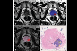
According to the preliminary findings, the readers spotted 31 lesions in 18 patients on mpMRI, 26 lesions in 16 patients with PSMA-PET, and 29 lesions in 18 patients on PET/MRI images. Of these, based on biopsy results, 21 of the lesions on PET/MRI were true positives, compared with 18 on mpMRI and 19 on PSMA-PET. A patient-level analysis showed that both mpMRI and PSMA-PET had a sensitivity of 92%, while PET/MRI had a sensitivity of 100%, Brembilla added.
Limitations of the study include the small number of patients so far and that a consensus has yet to be reached on how to report the PSMA-PET findings in a consistent way that would allow them to compare results across centers, he noted. The trial is ongoing and is expected to enroll 167 patients, he said.
“The combination of PSMA-PET and MRI could improve the detection of clinically significant cancer in clinically high-risk patients,” he concluded.