PET scans have visualized altered brain activity in individuals with alcohol use disorder (AUD), according to a recent study.
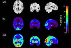
Researchers at Yale University in New Haven, CT, compared high-resolution brain scans between a control group and participants with AUD and observed lower synaptic density in the frontal cortex and striatum regions in those with AUD.
“Synaptic density deficits are evident, even in people with mild-to-moderate AUD, with greater deficits in those with greater drinking severity. These findings underscore the potential of synaptic restoration as a therapeutic target for AUD,” noted lead author Yasmin Zakiniaeiz, PhD, and colleagues. The study was published January 13 in the Journal of Clinical Investigation.
Alcohol use is a severe public health problem and is responsible for a substantial part of the global burden of disease and mortality. Preclinical studies in mice and rats have shown that chronic alcohol intake is associated with the loss of synapses in certain brain regions, such as the cerebellum and hippocampus. This loss of synapses is an early marker of neurodegeneration, the researchers explained. Yet there is a need to translate these preclinical findings to living humans, they noted.
To that end, the group recruited 32 people with AUD (17 women) and 29 controls (17 women). Individuals with AUD reported drinking on average five drinks per day, four days per week, for 12 years, while the control group (matched on sex, education level, and cigarette smoking) averaged less than one drink per day, less than one day per week, over 10 years.
All participants underwent scans in a high-resolution research tomograph brain PET system with a radiotracer (carbon-11 UCB-J), which binds to SV2A, a protein on synaptic vesicles. Levels of synaptic density were quantified by estimating the binding potential (BPND) of the tracer to SV2A across four regions of interest: the frontal cortex, striatum, hippocampus, and cerebellum.
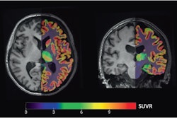
 Synaptic density images by Diagnostic Group. Parametric images of partial volume corrected BPND maps from two representative participants who were similar to the group mean.Journal of Clinical Investigation
“These findings suggest that the synaptic deficit in people with AUD may be dose-dependent, such that individuals who drink more could be ‘losing’ more presynaptic vesicles or synapses in these brain regions in a coordinated fashion,” the group wrote.
The researchers noted that due to the observational, case-control design of the study, it is not possible to determine whether the observed synaptic deficits are a consequence of AUD or a precursor of AUD and that future studies should use PET to determine the extent, if any, of synaptic density recovery with alcohol abstinence.
“This study translated preclinical findings to living humans with AUD, filling a crucial gap in the literature,” the group wrote.
The full study is available here.