F-18 FDG-PET/CT is superior to biopsies for detecting bone marrow involvement in patients with diffuse large B-cell lymphoma (DLBCL), researchers have reported.
The finding is from an analysis among newly diagnosed patients who underwent both bone marrow biopsy and PET/CT prior to any treatment initiation and could aid clinicians in making treatment decisions, noted first author Chunyan Yang, MD, of the Second Affiliated Hospital of Harbin Medical University in Harbin, China, and colleagues.
“Bone marrow biopsy has been regarded as the gold standard for assessing [bone marrow involvement] in DLBCL due to its ability to evaluate the bone marrow status of lymphoma patients. However, the advent and advancement of nuclear imaging technology have posed a challenge to its longstanding position in clinical practice,” the group wrote. The study was published December 15 in Radiation and Oncology.
DLBCL is an aggressive form of lymphoma and bone marrow involvement among patients is a critical prognostic indicator that significantly impacts disease staging and prognosis, the authors explained.
While bone marrow biopsy is the gold standard, it is an invasive procedure and carries the potential for patient anxiety and the risk of bleeding, they noted. Conversely, studies have suggested that F-18 FDG-PET/CT may replace biopsy based on its ability to detect bone involvement throughout the entire body, rather than at a single anatomical site, they added.
To further evaluate the potential role of PET/CT in these settings, the group compared outcomes among 34 patients ranging in age from 28 to 81 who underwent both biopsy and PET/CT imaging (Biograph 64 mCT, Siemens Healthineers) at their hospital in Harbin between 2017 and 2024.
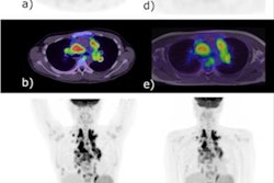
 The initial PET maximum-intensity projection images, and lymph nodes pathology confirmed DLBCL with normal (A), focal increased in the right humerus (B), diffuse increased (C, D).Radiation and Oncology
According to the analysis, PET/CT correctly identified 32 (56.1%) true-positive cases of bone marrow involvement, while bone marrow biopsy correctly identified 20 (35.1%) true-positive cases. Thus, PET/CT demonstrated superior accuracy (93% vs. 75.4%) and sensitivity (94.1% vs. 58.8%) compared with bone marrow biopsy, the researchers noted.
In addition, a survival analysis revealed a median progression-free survival (PFS) of 15 months and a two-year PFS rate of 47.9%. In a visual analysis, both a positive bone marrow biopsy (p = 0.016) and a positive PET/CT scan finding (p = 0.012) were significantly associated with shorter PFS.
“PET/CT played an important role in evaluating [bone marrow involvement] and predicting PFS in newly diagnosed DLBCL,” the group wrote.
In clinical practice, for newly diagnosed DLBCL patients, a PET/CT scan that is negative for bone marrow involvement could obviate the need for a bone marrow biopsy, unless there are apprehensions regarding potential oversight of low-grade lymphoma, the researchers suggested. Nevertheless, it has yet to be established that PET/CT can serve as a definitive substitute for bone marrow biopsy in clinical practice, they noted.
“Prospective large-scale studies are needed to further verify these results,” the group concluded.
The full study is available here.