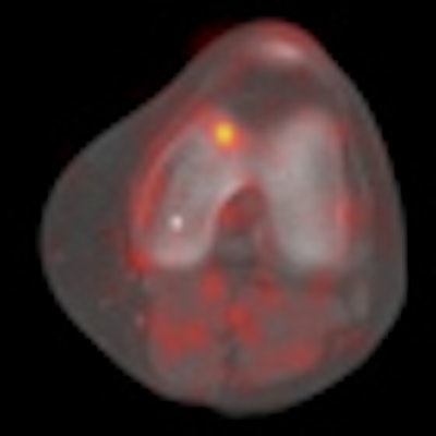
F-18 sodium fluoride (F-18 NaF) PET/CT may be a promising adjunctive technique to MRI for orthopedic applications, according to research from Stanford University in Palo Alto, CA.
The Stanford team found that the PET/CT technique can help detect additional abnormalities in 49% of patients with patellofemoral knee pain.
"Because F-18 sodium fluoride has such a high affinity for bone marrow, any process that increases the amount of exposed bone marrow will result in increased tracer uptake," said lead study author Christie Draper, PhD. "We hypothesize that F-18 sodium fluoride PET/CT can be used to visualize the localized regions of bone remodeling that occur in response to elevated tissue stress at the joint."
Draper presented the study results at the recent SNM annual meeting in Salt Lake City. The research received SNM's Correlative Imaging Council/Walter Wolf Award.
Patellofemoral pain forms behind the kneecap and accounts for approximately 25% of knee disorders in sports medicine clinics. MRI is often used as the gold standard to diagnose musculoskeletal abnormalities.
"Despite the incidents of this disorder," Draper said, "the specific mechanism of this pain remains unclear."
The researchers evaluated 22 subjects diagnosed with patellofemoral pain by a sports medicine physician. There were 12 males with a mean age of 31 years and 10 females with a mean age of 34 years. Enrolled patients experienced reproducible pain behind the kneecap during activities such as running, squatting, or going up and down stairs. Subjects were excluded if they had a history of other knee injuries or had prior knee surgery.
The researchers assessed pain during a physical examination by inducing pain in the patellofemoral joint and recording the location of maximum pain. F-18 NaF PET/CT scans (Discovery LS, GE Healthcare, Chalfont St. Giles, U.K.) were performed on all subjects' knees.
To minimize the effect of blood flow on tracer distribution, patients rested in a seated position for 30 minutes prior to injection of 5-10 mCi of F-18 NaF. The 22 enrollees continued to rest for another 60 minutes in a non-weight-bearing position prior to the PET/CT scans. They also received 3-tesla MRI scans to obtain axial and sagittal views.
The researchers also divided the knee into 26 anatomic regions for image analysis and to better localize increased tracer uptake. Two radiologists independently examined the images from all subjects, with PET images evaluated by a nuclear medicine radiologist.
Tracer uptake
The researchers discovered increased tracer uptake in 19 of the 22 patients with patellofemoral pain. The most common region for increased tracer uptake was the posterior patella in 17 cases and the femoral trochlea in five patients.
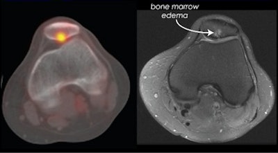
The researchers also compared regions of increased F-18 NaF uptake to see how the findings would correlate with cartilage damage as seen on MRI. The found that bone marrow edema, subchondral cysts, and cartilage damage were generally visible on both the PET/CT and MR images.
 |
| In the MR image (right), there is a region of bone marrow edema in the same location as increased tracer uptake seen at PET/CT (left). The images suggest that increased tracer uptake is related to the bone marrow edema. All images courtesy of Christie Draper, PhD. |
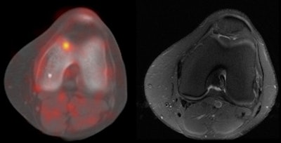
The group also found disagreement among PET/CT and MR images, as increased tracer uptake did not always correspond to structural defects. In one patient, increased tracer uptake was visible on the PET/CT image of the trochlea, but the MR image looked normal with no sign of cartilage or bone abnormalities.
 |
| Increased tracer uptake is visible on the side of the trochlea at PET/CT (left), while the MR image (right) shows the cartilage and bone intact. The images suggest there are regions of increased tracer uptake that do not correspond to structural defects using MRI. |
"This suggests there are a number of regions of increased tracer uptake in these patients that do not correspond to gross structural defects using MRI," Draper said.
In their final analysis, the researchers determined that 39% of the regions with increased tracer uptake corresponded to a similar finding on MRI. However, 49% of the regions did not correspond to anything on MRI. In 12% of the cases, bone or cartilage defects were seen on MRI but were not detected with F-18 NaF PET/CT.
Orthopedic imaging
Also, among the 22 subjects, 15 patients could localize their pain to either the medial or lateral side of the patellofemoral joint. Five patients identified the pain on the medial side of the joint, where there was increased tracer uptake. Another five patients identified pain on the lateral patellofemoral joint, with all five images showing increased tracer uptake in that location.
"This limited analysis suggests that pain may be related to increased tracer uptake in some subjects," Draper said. "This was limited by the number of patients studied and the resolution of the PET/CT images, but despite these limitations, F-18 sodium fluoride PET/CT shows some potential for diagnosing orthopedic conditions."
In conclusion, Draper noted that increased stress in the subchondral area causes pain in these patients and that condition could be one reason for the increased tracer uptake. "We hope to perform future studies comparing bone stress distribution with tracer distribution to evaluate this comparison," she added.
Draper also speculated that F-18 NaF PET/CT may help detect "metabolic abnormalities in the bone prior to the development of degenerative changes in the joint. We hope to perform long term follow-up studies on patients to evaluate this hypothesis."
By Wayne Forrest
AuntMinnie.com staff writer
June 22, 2010
Related Reading
F-18 NaF PET/CT can help with atherosclerotic plaque detection, June 11, 2010
CMS OKs F-18 NaF imaging coverage, March 8, 2010
F-18 fluoride PET/CT may better manage painful bone metastases, November 29, 2009
Sodium fluoride PET/CT changes management of foot pain patients, November 3, 2009
1T extremity MRI accurately shows knee disorders, June 24, 2009
Copyright © 2010 AuntMinnie.com


