CHICAGO - The management of atherosclerosis, the No. 1 cause of death in the U.S., has been reinvented by advances in imaging technology, according to a panel presentation on Sunday at the RSNA 2011 meeting.
Led by Dr. Geoffrey Rubin, radiology chair at Duke University Medical Center, and Dr. Zahi Fayad, PhD, professor of radiology and cardiology at Mount Sinai Medical Center, the opening-day panel explored the evolution of CT angiography (CTA) as the leading imaging modality for atherosclerotic disease, as well as the growing contributions of other modalities in the refinement of atherosclerosis care.
"What led me into cardiovascular imaging and through the development of CT angiography was an appreciation of the beauty and profound information that is contained within the images that we acquire," Rubin said. Owing to its enormous flexibility and diagnostic power, CTA has become the leading modality in cardiovascular disease management. "CT angiography allows us to volumetrically look at complex relationships of blood vessels and glean important and subtle abnormalities," he said.
In a talk titled "Coronary CT Angiography -- 20 Years Old and All Grown Up," Rubin explained how the modality wasn't always so flexible, and the images not so exquisitely detailed. Rubin's first CT angiography exam in 1991, a renal artery study acquired on a single-detector-row helical scanner, was a blurred abstraction compared to today's volumetric images, but it accurately demonstrated both the relevant anatomy and the pathology -- capabilities that improved greatly with the introduction of four-detector-row scanners in 1998 and continued through the development of today's volumetric scanners. Diagnoses that had remained stubbornly elusive at conventional angiography suddenly became clear, and were available in a 3D display that could be easily shown and transmitted to referring physicians, he said.
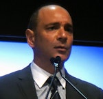
 Dr. Geoffrey Rubin.
"The introduction of dynamic capability with CTA and, in particular, wide area detectors, allowing us to simultaneously look at blood flow volumetrically, has added further to the tremendous capabilities of this technique," Rubin said. In vascular imaging, understanding of acute aortic syndromes and aortic dissection has been revolutionized by the ability to examine blood flow, which has revealed the dynamics of the intimal flap and its relationship to cardiac blood flow, and imparted new understanding to atrial-septal disease.
The introduction of MDCT heralded an age of broader anatomic coverage and fine spatial resolution that has continued in the years since, simplifying clinical management and replacing a wide array of invasive techniques or improving surgical planning. Today, "virtually every vascular bed is evaluated routinely with CT angiography," he said. But the technique is still advancing, and more work needs to be done in key areas.
Endovascular device selection and characterization is one such area. During the development of stent grafting techniques, experts told him that CT would never replace conventional angiography, an area where it is now the mainstay, Rubin said.
Ultrasound was traditionally used for measuring the aorta, though it couldn't match the accuracy of volumetric CT, which produced true orthogonal representation of the cross-section of the aorta, leading to the first accurate way to size stent grafts.
Also key to vascular CT's development were techniques developed for continuous measurement of vascular dimension by calculating mean diameter from cross-sectional area. These developments by Rubin and colleagues led to commercial implementations on workstations that are now the standard, he said. For endograft assessment, CT has also "stepped up in a big way to show us the detail," ultimately leading to design improvements in the stents themselves. A new technique, transcatheter aortic valve implantation, was made possible by improvements in endograft measurement and assessment and, via two manufacturers of the replacement valves, is set to revolutionize the treatment of aortic valve stenosis.
"I hope you are looking into these techniques in your hospital," Rubin said. "It is a very exciting area for radiologists to involve themselves in, and a very important one."
Also emerging as a key technique for triaging patients with positive CTA results is fractional flow reserve (FFR), which measures blood flow based on volumetric CT data. FFR "has a tremendous predictive value in determining what types of coronary lesions will ultimately result in major myocardial infarction or death," he said. Recently published two-year results from the Fractional Flow Reserve Versus Angiography for Multivessel Evaluation (FAME) trial by Tonino and colleagues showed a 33% reduction in the risk of death or major cardiac events when percutaneous coronary angioplasty and stenting was based upon fractional flow reserve measurements at CTA versus angiography, Rubin said.
Also good news but a little disconcerting, he said, is that correlation of lesions at coronary CTA with fractional flow reserve shows that "75% of those lesions turn out to be false positive," meaning that they do not have a low enough FFR to cause ischemia -- and that coronary CTA alone can't distinguish them.
This understanding has led to the application of computational fluid dynamics to coronary CTA data based on "a tool used every day in characterizing flow around structures such as jet aircraft or race cars in order to understand how a structure interacts with flowing material," Rubin said. Calculations based on this model allow for continuous mapping of FFR values, which enable a characterization of the coronary tree not possible from images alone, he said.
True to the earlier FFR revelations, a recent study shows that application of this model has a striking impact on the specificity of coronary CTA, raising specificity from 25% to 82%. The technique shows great promise in reducing false positives in CTA, he said.
 |
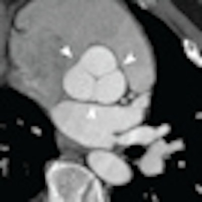
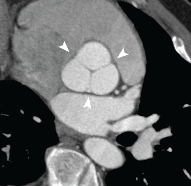
| Normal aortic root at coronary CTA illustrating the complex cornet shape above the basal ring. Image courtesy of Dr. Geoffrey Rubin. |
Finally, the traditional focus on arterial lesions has led to new tools to map end-organ perfusion. "It is ultimately through the [blood] supply and demand to the end organ through perfusion, as well as the demand to the end organ through metabolism, that we will really understand the relevance of these vascular lesions," Rubin said. In practical terms, for patients presenting with substernal chest pain, it's not always clear whether it is the CTA-detected vascular lesions that are causing the pain.
"The availability of techniques now that allow wide area coverage through either shuttle mode of the CT or high-pitch mode or wide area detectors allow for CT perfusion studies to be performed," he said. In patients with multiple possible culprit lesions, perfusion pinpoints the cause of ischemia and is well-correlated to MR and SPECT perfusion studies, he said. Extending the study to look at delayed enhancement can distinguish true myocardial infarction from recoverable myocardium.
Along with the great advances for CTA have come strident reminders of the dangers through the lay press, and some highly popularized cases of patient harm, he said. "But I am very excited that the developments coming down the line ... are all serving to substantially reduce the radiation exposure of CT angiography," Rubin concluded. "I strongly believe that in the future the vast majority of CT angiographic procedures will be performed at less than 1 mSv."
Multimodality atherosclerotic imaging
In his talk about "bleeding-edge imaging and therapy in vascular disease," Fayad highlighted developments in multimodality atherosclerosis imaging, prevention, diagnosis, and treatment. Atherosclerosis is the most important challenge among four noncommunicable diseases -- cardiovascular disease, diabetes, cancer, and chronic respiratory disease -- that are challenging health, finance, and socioeconomic well-being in the 21st century, he said.
Early detection of risk factors, such as hypertension for cardiovascular disease, will play a major role, he said. That means more attention to diet and physical activity. Reduction in tobacco use, excessive alcohol consumption, etc., will be important for reducing the toll, as will accurate diagnosis focusing on imaging, Fayad said.
"When we talk about atherosclerosis, we think most of the time of the coronary arteries, but we know that atherosclerosis is a disease that burdens the whole body," Fayad said. "It can affect the carotid arteries, which leads to stroke and peripheral disease," sharing many characteristics with coronary artery disease. "Nonsignificant" luminal stenosis of less than 50% is really the main culprit in acute myocardial infarction, "and there's more and more evidence that it's the nonstenotic lesions that lead to stroke," he said.
Investigations in lesion pathology have led to characterization of two main types of atherosclerotic lesions: Stable plaque -- characterized by the presence of thick fibrous cap, a modest lipid pool, and few inflammatory cells -- is distinguished from the more dangerous unstable plaques, characterized by a thin fibrous cap, low collagen content, large lipid pool, many inflammatory cells, and angiogenesis.
 Dr. Zahi Fayad, PhD.
"These are the features that you have to use imaging to try to detect," he said. And since these unstable lesions are nonstenotic, a typical CT angiography study won't characterize them as significant.
Because drug therapy is the principal method of mitigating disease progression, improving drug development in the pharmaceutical industry presents another major challenge, Fayad said. More and more drugs are needed at the same time that fewer are being approved by the U.S. Food and Drug Administration (FDA).
Currently, the drug development cycle is very long and the need for money is enormous, he said. Accepting or rejecting drugs earlier in the approval process -- at phase II, for example -- is one step that will accelerate drug development. The use of cardiovascular imaging biomarkers in the assessment of atherosclerosis is one idea that has gained some acceptance in both the FDA and the European Medicines Agency.
The experience with cancer provides some insight -- FDG uptake after treatment is predictive of the future course of prostate cancer, for example -- and now the idea has shown applicability to vascular imaging. At this year's RSNA meeting, Tawakol, Fayad, and colleagues will present results from the first multicenter trial of statins showing the impact of different dose levels on FDG uptake post-treatment, he said.
"There are a whole array of imaging tools that are beginning to be applied to the imaging and treatment of atherosclerosis," Fayad said.
Even "plain vanilla" MRI is useful for identifying plaque and characterizing the burden of disease, but new techniques are taking the modality further, he said. For example, multiweighted MRI images can help identify the composition of the plaque to convey a sense of the lipid-rich necrotic core with or without contrast. T2-weighted MR images can identify the cholesterol deposits due to their hypodensity, along with the calcifications. One step further, dynamic contrast-enhanced MR images of the type used for tumor imaging can be used to identify angiogenesis of inflammation, he said.
Already tried in humans is MRI using iron oxide contrast with microphages, and fibrin-specific MRI contrast, though the latter agent never made it to market due to economic constraints.
FDG uptake shows vascular inflammation
Vascular imaging using FDG-PET to measure glycolysis is one approach for creating contrast. It's an important approach because high-risk plaque has a high level of activated macrophages and, therefore, a high level of FDG activity and glucose uptake, Fayad said. "This technique can be used today to look at vascular inflammation in a multitude of inflammatory diseases, and we and others have been looking at it to use it as a biomarker of inflammation measurement."
Techniques using standard FDG have been validated in vivo by histopathology in carotid atherectomy patients, he said. In a multicenter study not yet published, FDG was able to distinguish between two groups of patients -- randomized to high-dose or low-dose statins -- to measure significant differences in FDG uptake levels after treatment. The trial is fundamental to understanding the power of this imaging test, he said.
Whether this knowledge will predict atherosclerotic events is one of the most important questions to be answered in terms of the clinical applications, according to Fayad. But there are hints that it works, from a trial of cancer patients that studied FDG uptake in tumors. Those patients with higher levels of vascular inflammation per FDG had more atherosclerotic events, he said. And new non-FDG radiotracers, such as carbon-11 PK11195, found on many macrophages, are also showing promise, distinguishing symptomatic from asymptomatic patients in a recent study.
MRI plus FDG
Merging FDG uptake with MRI in an integrated PET/MR scanner adds another dimension, Fayad said, regarding another recent study undertaken by his group in "PET-weighted imaging." Why perform simultaneous PET and MRI for vascular imaging?
"We wanted to focus on being noninvasive," he said. "We wanted a technique that was highly reproducible because we're interested in quantification. We wanted a technique that can give us a sense of plaque composition and burden in combination with metabolism and plaque information."
The group's multicenter clinical trial showed that it was "robust and useful" for characterizing atherogenesis, according to Fayad. Twelve months after initiation of niacin therapy, the multimodality approach clearly demonstrated disease regression.
MRI is being used more and more in large, population-based MRI studies, including the Framingham Heart Study, the Multiethnic Study of Atherosclerosis (MESA), and the Rotterdam Heart Study, where MRI is being used to "try to identify the known correlation of atherosclerosis risk factors, as well as imaging-based atherosclerosis assessment," Fayad said.
Finally, a multicenter study published last month showed the effectiveness of the atherosclerosis drug dalcetrapib using FDG-PET and MRI in combination. PET/MRI scanning is going to have a big impact in understanding atherosclerosis while protecting patients from radiation exposure, Fayad said. The recently FDA-approved Avalanche photodiode PET/MR scanner (Philips Healthcare) is another promising addition to the modality.
CT in living color
Color is another addition in atherosclerosis imaging, enabled by the development of energy-resolving detectors for spectral CT scanning. "I think we're going to finally have the potential to utilize CT to do better imaging," he said.
One prototype detector has eight levels of energy distribution and has shown it can distinguish gold from iodine for better tissue characterization. A few spectral scanners are available now for animals, and models may be available for use in humans in the near future. "We feel that detectors like these can characterize the tissue without need of precontrast imaging, so that's going to be important," wielding a big impact on clinical radiology by reducing dose substantially, he said.
Nanotechnology
Nanotechnology presents a great opportunity to combine drug delivery and imaging of atherosclerosis. "This could be a way to detect disease before health has deteriorated," he said. Nanotechnology can be used to create sensors, or imaging tools based on nanoparticles. It can deliver and monitor the effect of therapeutic agents, and represents an excellent way to improve the understanding of disease and apply it later in the clinical realm. Combined nanotechnology and therapy is the latest frontier for this research, he said.
The field was initially driven by cancer research, but there are new investments at the National Institute of Health's National Heart, Lung, and Blood Institute to drive research in the field, he said.
"We're seeing a huge interest in the number of publications and interest in molecular imaging," Fayad said. Most of these techniques are applied in the preclinical setting, he said, but look for the emergence of one of these agents soon in the clinical realm.
One example of a nanoparticle mimicking biology -- always a good bet, he noted -- is the use of a high-density lipoprotein as a contrast and drug-delivery platform that can be used with MRI, PET, and CT, while incorporating a drug at the core of the lipoprotein for local delivery of therapy. Currently, many oncology applications for nanotechnology are in clinical use, and several companies are "close to filing [investigational new drug] applications with the FDA for nanotechnology products," he said.
And what starts in cancer can break through into vascular disease care. Fayad's group is working on a "potent anti-inflammatory" agent that calmed arterial inflammation in a rabbit model within a week of injection, and is now being applied in human subjects.
Along with these developments, "I'm very excited about new developments in PET/MR, as well as spectral CT," as well as new therapeutic applications of nanoparticle agents that are "useful for both diagnosis and therapy," Fayad said.


