X-ray fluoroscopic guidance with iodinated contrast for endovascular procedures poses well known risks, including that of radiation exposure to both patient and physician. A search for safer methods led researchers in Wisconsin to look at MRI as an alternative. Writing in the September Journal of Vascular and Interventional Radiology, they concluded that as MRI technology evolves, it could compete with fluoroscopy to offer safer image-guidance techniques for endovascular procedures.
For endovascular MR image guidance to be feasible, catheters must be both visible and compatible with the MR imaging environment, wrote study authors Dr. Reed Omary, Orhan Unal, PhD, and colleagues from the University of Wisconsin Medical School in Madison (JVIR, Sept. 2000; Vol.11:9, pp. 1079-1085).
Catheter visualization with MRI has traditionally been based on either active or passive techniques. Active methods require the placement of one or more small radiofrequency coils at the end of a catheter. The catheter position is obtained from the position of the MRI signal detected from the coil. Most methods for passive catheter visualization rely on artifacts induced by differences in magnetic susceptibility between the catheter and the surrounding tissues, the authors wrote.
An alternative technique developed by the Wisconsin researchers utilizes the T1-shortening effect of gadolinium (Gd)-based contrast agents, which depict the catheter as a positive rather than a negative signal. To track the catheter as it moves, they created a real-time MRI tracking system to reconstruct and display images as rapidly as the data is acquired.
Real-time imaging
The researchers used an MR imaging pulse sequence with high temporal and spatial resolution for passive catheter tracking on a cardiovascular 1.5-tesla MR scanner with EchoSpeed gradients. (GE Medical Systems, Waukesha, WI).
The echo sequence was two-dimensional, radiofrequency-spoiled and gradient-recalled. It incorporated time-resolved imaging of contrast kinetic elements, temporal interpolation, view sharing, and zero-filling, with a projection dephaser in the slice direction, the authors wrote.
Images were acquired with the aid of a quadrature head coil, and reconstructed on an UltraSparc II workstation (Sun Microsystems, Mountain View, CA).
The optimal gadolinium concentration was determined by using gadopentetate dimeglumine (Magnevist; Berlex Laboratories, Wayne NJ) as the Gd chelate and diluting it with saline. Six Gd solutions, ranging in concentration from 2% Gd to 12% Gd, were placed in 5-F dilators and used as catheters in the experiments.
Tracking Gd catheters
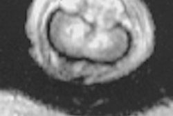
The catheters were placed vertically, open end down, in a glass beaker filled with nonfat yogurt used to mimic tissue. Imaging was then performed both with and without a projection dephaser.
The team measured signal-to-noise ratio, contrast-to-noise ratio, and enhancement ratio to determine the optimal Gd concentration for catheter depiction. Signal measurements (in each catheter, in the yogurt adjacent to the catheter, and in the air outside the phantom) were determined by averaging region-of-interest values from 50 images.
About 275 images were acquired during each set of catheter movements.
The results showed that peak catheter visibility occurred with 4% to 6% Gd concentrations, and the catheters were more visible when the projection dephaser was used.
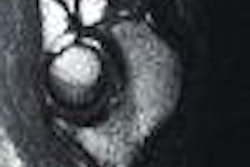
In order to test tip tracking, a single-lumen, 5-F catheter with a 4% Gd solution was marked with seven stripes 2-12 mm apart, and moved into and out of an acrylic phantom with channels (to mimic blood vessels). Tip tracking was accurate within ± 0.41mm as determined by the root mean square method -- well within the authors' definition of ± 1.0 mm as a clinically permissible error.
The experiments performed in static phantoms were just the first step, the authors noted, recommending that the methods be verified in animals to confirm safety. The authors noted that better spatial and temporal resolution will be needed before the method can compete successfully with x-ray fluoroscopy.
While significant advances in software and hardware will be needed before the technique can be performed safely in humans, the initial results are encouraging, the group concluded.
"This technique should be applicable to many vascular territories and may be useful in the development of future MR imaging-guided endovascular procedures," they wrote.
By Jonathan S. Batchelor
AuntMinnie.com staff writer
September 28, 2000
Let AuntMinnie.com know what you think about this story.
Copyright © 2000 AuntMinnie.com


