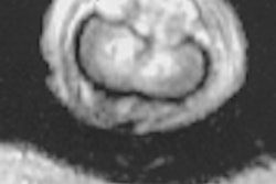
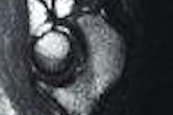
Does adding a little FLAIR to contrast-enhanced MRI improve the depiction of leptomeningeal metastases in the brain? Not really, according to researchers at the MD Anderson Cancer Center, who compared contrast-enhanced T1-weighted MR to fluid-attenuated inversion-recovery images.
FLAIR sequences have been a routine part of brain imaging at the Houston hospital since 1997, wrote Dr. Sanjay Singh and colleagues in the October issue of Radiology.
But does it offer neuroradiologists any more information than they could have gathered without it?
"Imaging of leptomeningeal metastases is important since cerebrospinal fluid cytologic examination results are often falsely negative and leptomeningeal metastases occasionally may be asymptomatic and unsuspected clinically," they said. "This has prompted the search for more sensitive imaging techniques" (Radiology, October 2000, Vol.217:1, pp.50-53).
In a retrospective analysis, the group pulled the records of 58 patients who underwent both FLAIR and contrast-enhanced T1-weighted spin-echo MRI. The exams were performed with a 1.5-tesla system (Signa Horizon, GE Medical Systems,Waukesha, WI). The T1-weighted images were obtained in all three planes after an intravenous injection of gadopentetate dimeglumine (Magnevist, Berlex Imaging, Wayne, NJ).
The FLAIR sequences were in the transverse plane with flow compensation and were fast sequences: 10,000 repetition time msec/147echo time msec/2,200 inversion time msec. The echo train length was 22.
A neuroradiologist who knew of the malignant findings in all patients reviewed the transverse FLAIR sequences, assigning a rating of positive, indeterminate, or negative for leptomeningeal metastases, the authors explained.
Out of 58 cases with FLAIR imaging, the reader assigned 20 as positive, 32 as negative, and indeterminate in six cases. FLAIR had an overall sensitivity of 34%. For the contrast-enhanced T1-weighted images without FLAIR, the reader pegged 38 cases as positive for leptomeningeal metastases, 17 as negative, and three as indeterminate. The sensitivity for this technique was 66%, the group reported.
In three instances, leptomeningeal metastases were detected only by the FLAIR images. These lesions were located in the cerebral sulci. However, 20 metastases could only be detected with contrast-enhanced T1-weighted imaging. These findings were located either in the cerebral sulci or in cisternal spaces.
"Our data indicate that, at least for leptomeningeal metastases, FLAIR images are not better than contrast-enhanced T1-weighted images. Although there are cases in which FLAIR images depict abnormalities not identified on contrast-enhanced T1-weighted spin-echo images, overall the latter are more sensitive," the paper stated.
The group acknowledged that its study did have some limitations: Because of 100% disease prevalence in this patient population, the specificity of the two techniques could not be determined. In addition, because only one reviewer was used, there was no assessment of interobserver reliability.
Still, while FLAIR imaging shows promise, it currently has limited value for the assessment of malignant brain metastases, they concluded.
By Shalmali PalAuntMinnie.com staff writer
October 19, 2000
Let AuntMinnie.com know what you think about this story.


