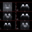
(Radiology Review) A recent study by Italian researchers at the University of Pisa found that contrast-enhanced 3-D MR angiography (MRA) was an excellent method of evaluating carotid artery disease. Presently, digital subtraction angiograpy (DSA) is the gold standard for classifying carotid arterial stenoses, but carries a 1% risk of stroke. Other noninvasive methods are used preoperatively and include Doppler ultrasound, spiral CT, and MRA.
According to the group, conventional MRA tends to overestimate high-grade stenoses because turbulent flow causes intravoxel phase dispersion, which is overcome by using contrast-enhancement.
"The introduction of high-performance gradients allowed rapid acquisition in an attempt to image the first passage of paramagnetic contrast media in the arteries," they wrote in their study published in Stroke.
The object of studying 92 patients with suspected carotid disease was to assess the diagnostic exactness of contrast-enhanced MRA (CEMRA), and to evaluate the misclassification rate.
A Horizon 1.5-tesla system was used (GE Medical Systems, Waukesha, WI). Images were obtained with a 3-D fast spoiled gradient echo acquisition in the oblique coronal plane using a dedicated phased-array neovascular coil. An IV injection of 30mLs of Gd-DTPA was administered using a power injector at 2mL/s. Three-dimensional reconstruction was performed using a dedicated workstation. ICA luminal narrowing was independently classified by two radiologists into four groups: <50%, 50-70%, >70%, and occlusion.
Comparing CEMRA with DSA, results demonstrated a sensitivity of 97%, a specificity of 82% and an accuracy of 92.5%. Five of six subtotal occlusions (string sign) were demonstrated on CEMRA and 15 of 17 ulcerated ICA plaques were correctly identified on CEMRA.
Carotid endarterectomy, ICA stent placement, and PTA were used to achieve revascularization in 48 patients. These treatments were considered necessary for patients with >70% ICA stenosis, and in symptomatic patients with a 50-70% stenosis. CEMRA misclassified six patients. One patient was thought to have an occlusion that was a subtotal occlusion on DSA, and five patients were overestimated as >70% on CEMRA, but were considered 50-70 % on DSA.
The authors concluded "the high diagnostic accuracy and limited misclassification rate suggest that CEMRA can be considered a powerful tool for the preoperative, noninvasive evaluation of atherosclerotic pathology of carotid arteries."
Contrast-enhanced three-dimensional magnetic resonance angiography of atherosclerotic internal carotid stenosis as the noninvasive imaging modality in revascularization decision making Cosottini, M., et. al.Department of neuroscience, University of Pisa, Pisa, Italy
Stroke 2003 March; 34:660-664
By Radiology Review
August 14, 2003
Copyright © 2003 AuntMinnie.com


.fFmgij6Hin.png?auto=compress%2Cformat&fit=crop&h=100&q=70&w=100)


.fFmgij6Hin.png?auto=compress%2Cformat&fit=crop&h=167&q=70&w=250)


