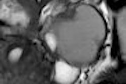
(Radiology Review) MRI T1-weighted hypointense lesions on unenhanced in-phase images and T2-weighted hyperintensity or arterial hypervascularity are highly suggestive of malignancy in patients with liver cirrhosis, according to a study published in the American Journal of Roentgenology.
Radiologist Dr. Jeong-Sik Yu and colleagues from Yonsei University College of Medicine in Seoul, South Korea, wrote that, in the course of hepatocarcinogenesis, premalignant lesions can undergo fatty change preceding hepatocellular carcinoma.
To determine the predictive value of MRI for diagnosing malignancy in fat-containing nodules in the cirrhotic liver, 38 patients with liver cirrhosis and focal lesions containing fat, found on chemical shift gradient-echo MRI, underwent further investigation.
Patients with numerous 5-mm or larger fat-containing nodules were divided into two groups -- group A patients had up to four lesions, while group B patients had more than 10 lesions. "Positive predictive values (PPVs) for benignity and malignancy were calculated on the basis of lesion size, T1-weighted hypointensity, T2-weighted hyperintensity, and arterial hypervascularity on the initial MR images," they wrote (AJR, April 2007, Vol. 188:4, pp. 1009-1016).
For patients with up to four lesions (group A), 47% of lesions became malignant. Mean diameter was 18.8 mm for malignant lesions compared with a mean diameter of 10.5 mm for benign lesions. For patients with 10 lesions or more (group B), all lesions were benign.
"The PPV of larger (≥ 15 mm) fat-containing nodules for malignancy was 85%. Six (55%) of 11 immediately diagnosed hepatocellular carcinomas were entirely hypointense on unenhanced in-phase T1-weighted images," they said. For predicting malignancy, they found a 100% PPV for T2-weighted hyperintensity and arterial hypervascularity in patients with up to four lesions.
The authors concluded that for patients with liver cirrhosis, a finding of larger lesions (≥ 15 mm) with T1-weighted hypointensity on in-phase images indicated malignancy of the fat-containing nodules. When multiple small lesions (< 1 cm) are shown, benign disease is indicated, they stated.
"Fat-Containing Nodules in the Cirrhotic Liver: Chemical Shift MRI Features and Clinical Implications"
Jeong-Sik Yu el al
Department of Radiology, Yonsei University College of Medicine, YongDong Severance Hospital, 146-92 Dogok-Dong, Gangnam-Gu, Seoul 135-720, South Korea
AJR 2007 (April); 188:1009-1016
By Radiology Review
June 13, 2007
Copyright © 2007 AuntMinnie.com


