MRI's ability to find abnormalities in severe cases of hypoglycemia proved beneficial to Singapore physicians in diagnosing patients who consumed an erectile dysfunction drug that's illegal in that country ... and that caused several patient deaths.
At the heart of the situation is Power 1 Walnut, an herbal preparation adulterated with the synthetic drug sildenafil, the active ingredient in the erectile dysfunction drug Viagra, which is produced by Pfizer of New York City.
Between January and March 2008, four Singapore hospitals reported an increase in the number of individuals without diabetes coming to their facilities with hypoglycemic symptoms. Without treatment, severe hypoglycemia is potentially fatal and can cause a loss of mental acuity.
The patients thought they were taking a natural medicinal imitation of Viagra, according to a study performed by researchers from the National Neuroscience Institute in Singapore and published in the January issue of Radiology (January 2009, Vol. 250:1, pp. 193-201). However, further analysis of the product by study co-author Dr. C.C. Tchoyoson Lim and colleagues uncovered high amounts of glibenclamide, an oral hypoglycemic agent used to treat diabetes. Physicians were able to connect the consumption of Power 1 Walnut to the cases.
Suspected cases
After a review of clinical and MRI records of 76 patients in the National Neuroscience Institute and four Singapore general hospitals (Tan Tock Seng Hospital, Changi General Hospital, Singapore General Hospital, and National University Hospital), the researchers narrowed their study to eight cases of suspected severe hypoglycemia of unknown origin between January 1 and March 31, 2008.
The eight patients, all men between the ages of 26 and 73 years, were admitted for focal neurologic deficit, seizures, altered sensorium, and/or coma. They had no history of diabetes and showed positive blood toxicology results for glibenclamide. Two patients were confirmed to have taken the illegal sexual enhancement product, and the other six men were suspected of the consumption.
The procedures included either 1.5-tesla or 3-tesla MRI scans between the second and ninth days of admission, as well as diffusion-weighted imaging and, in some patients, MR angiography, dynamic contrast-enhanced perfusion MR imaging, and MR spectroscopy.
Severe hypoglycemia
In cases of severe hypoglycemia, MRI has tended to show abnormal findings, typically in the cerebral cortex, hippocampus, and basal ganglia bilaterally. However, the authors wrote, "these MR imaging studies in hypoglycemia are usually confined to case reports in patients with diabetes who had accidentally overdosed while receiving treatment with oral hypoglycemic agents."
Time-of-flight MR angiography of the intracranial circulation was also performed in some patients. In addition, first-pass dynamic susceptibility contrast-enhanced perfusion MR imaging and proton MR spectroscopy were performed on a 1.5-tesla MRI scanner (Signa Excite, GE Healthcare, Chalfont St. Giles, U.K.) in one patient.
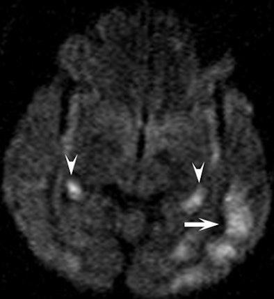
In a review of the images, the researchers found the hippocampus was affected bilaterally in seven patients, with variable involvement of the cortex in the parietal lobe (seven patients), temporal lobe (six patients), occipital lobe (five patients), and frontal lobe (five patients).
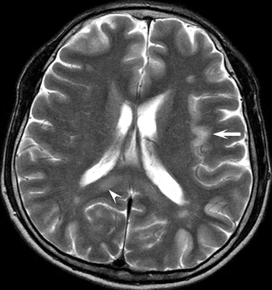
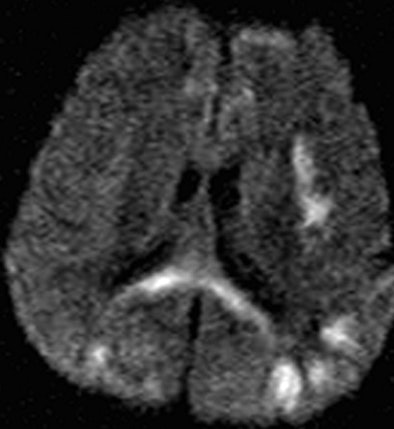
In seven patients, there was cortical involvement that the authors described as "patchy noncontiguous" in four cases and "confluent" in three patients, with sparing of the subcortical white matter and cerebellum. Three patients displayed abnormalities of the splenium of the corpus callosum in addition to the cortex.
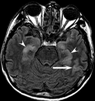 |
| Clinical images of a 51-year-old man found unconscious with a Glasgow Coma Scale score of 7 and withdrawal to pain. Above image shows fluid-attenuated inversion recovery, while below is a diffusion-weighted image that shows an increase in signal intensity in the head, body, and tail of the hippocampus bilaterally (arrowheads) and in the cerebral cortex (arrow). All images courtesy of Radiology and the National Neuroscience Institute. |
 |
 |
|
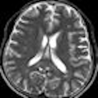
Above is a T2-weighted MR image of bilateral patchy hyperintense lesions in the cerebral cortex, including the insula (arrow). There is also involvement of the splenium of the corpus callosum (arrowhead). On the corresponding diffusion-weighted MR image below, the hyperintense lesions are more prominent than above. |
 |
"These findings are consistent with those of previous studies that described selective vulnerability of the brain to hypoglycemic damage and characteristic lesion distribution," the authors wrote.
Patient outcomes
As for the patients, the overall outcomes were not good. Within two weeks of admission, one patient had recovered completely, one man developed a slight disability, and five patients had moderate to severe disability. One patient died of complications after 28 days in a persistent vegetative state.
By the time the study was finalized late last year, five patients had died or developed a pathological condition. "Although clinical outcome has been correlated with the severity and duration of hypoglycemia," the authors wrote, "the utility of neuroimaging findings for prognosis has not yet been established. Further analysis remains to be performed."
The authors also recommended that as more data emerges from these cases "and criminal investigations continue, radiologists and clinicians should be aware of the MR imaging features of severe hypoglycemia."
In patients who have taken sexual enhancement products containing glibenclamide, the ability of diffusion-weighted MRI to discover cortical and hippocampal abnormalities, with additional involvement of the corpus callosum and internal capsule in some cases, can be most beneficial, the authors concluded.
By Wayne Forrest
AuntMinnie.com staff writer
February 25, 2009
Related Reading
PET becomes a godsend for newborns with hyperinsulinism, August 7, 2007
Hyperglycemia associated with increased cancer risk, March 16, 2007
Sexual healing: Imaging takes a roll in the hay with human relations, April 29, 2004
Copyright © 2009 AuntMinnie.com


