Sunday, November 29 | 11:35 a.m.-11:45 a.m. | SSA14-06 | Room E450A
Researchers at the Medical University of Vienna are using 3-tesla MRI to assess the structure and function of peripheral nerves. Their study shows that advanced 3-tesla imaging methods, such as diffusion tensor imaging (DTI) and tractography, could add valuable information to the treatment of various peripheral nerve conditions."The higher field strength and better signal-to-noise ratio allows the application of diffusion tensor sequences with a comparably high resolution in a clinically acceptable imaging time of six minutes," said lead author Dr. Gregor Kasprian in the University's department of musculoskeletal and neuroradiology. "Using 3T, surface and/or dedicated wrist coils, even fascicular structures of peripheral nerves can be three-dimensionally assessed."
The study evaluated 13 patients with localized upper extremity nerve lesions, which included cubital tunnel syndrome and carpal tunnel syndrome. Researchers included six normal patients for the control group. All were imaged using a 3-tesla MRI scanner and an axial echo-planar single-shot DTI-weighted sequence coregistered with short-tau inversion recovery (STIR) and T2-weighted sequences.
"Diffusion tensor imaging not only allows for the assessment of nerve structure and the 3D anatomy, it also provides quantitative data on the functional integrity of peripheral nerves in various conditions -- nerve compression syndromes, traumatic nerve lesions, plexus lesions, and nerve tumors," Kasprian said. "Animal studies recently demonstrated that this technique can noninvasively provide data on nerve regrowth and regeneration."
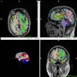
Upon further analysis, the study found that 3-tesla MRI with diffusion tensor imaging allowed for the microstructural characterization of 3D anatomy and pathology of upper extremity nerves. By 3D visualization of the brachial plexus nerve roots, tractography was helpful in preoperative surgical planning.
"Peripheral nerve tractography provides noninvasive quantitative and structural data, which can be used to assess peripheral nerve pathology with a higher sensitivity and especially with a higher specificity than the usual methods of MR neurography and ultrasound," Kasprian said. "The exact location and severity of peripheral nerve damage can be evaluated."
In the future, he added, the technique may be applicable in various clinical conditions and could aid in nerve infiltration by soft-tissue tumors, monitoring of nerve regeneration, planning of nerve surgery, and validation of surgical methods.


.fFmgij6Hin.png?auto=compress%2Cformat&fit=crop&h=100&q=70&w=100)



.fFmgij6Hin.png?auto=compress%2Cformat&fit=crop&h=167&q=70&w=250)


