Sunday, November 29 | 11:35 a.m.-11:45 a.m. | SSA16-06 | Room N228
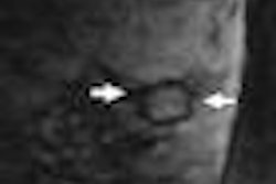
Researchers at M. D. Anderson Cancer Center have found that T1 fluid-attenuated inversion recovery (FLAIR) postcontrast imaging of the spine can significantly improve cerebrospinal fluid suppression and increases the conspicuity of bone lesions, disk herniations, and epidural metastasis.In the study, a musculoskeletal radiologist and a neuroradiologist reviewed contrast-enhanced MRI of the thoracic and lumbar spine with a 3-tesla scanner in 156 patients. All patients were imaged with sagittal postcontrast T1 spin-echo (SE) and T1 FLAIR sequences.
The radiologists then compared the sequences in each category, evaluating FLAIR or SE results. The categories were bone lesion, disk herniation, other epidural disease, and cerebrospinal fluid suppression.
The radiologists agreed that cerebrospinal fluid-cord distinction was significantly better with T1 FLAIR, and bone lesions were slightly more conspicuous on T1 FLAIR. In addition, they concurred that disk herniation was slightly more conspicuous on T1 FLAIR. Other epidural lesions also were deemed slightly more conspicuous on T1 FLAIR.
"In our study, T1 FLAIR slightly increased the conspicuity of metastases in the bone marrow, and no metastases seen by T1 SE were missed by T1 FLAIR imaging," said lead author Dr. Komal Shah, assistant professor in diagnostic imaging at M. D. Anderson Cancer Center at the University of Texas in Houston.
At 1.5 tesla, Shah said spin-echo T1 sequences of the spine show the cord well against dark cerebrospinal fluid. "However, at 3T," he added, "the spin-echo T1 sequence results in cerebrospinal fluid signal that is much closer to cord signal. T1 FLAIR can be used to improve the distinction between cerebrospinal fluid and cord at 3T by suppressing the signal from fluid."


