Coupling an MRI contrast agent to a nanodiamond can produce "dramatically enhanced" signal intensity and "vivid image contrast," according to a new Northwestern University study published in Nano Letters.
Researchers say the technique paves the way for the clinical use of nanodiamonds to deliver therapeutics and remotely track the activity and location of the drugs.
Thomas J. Meade, Ph.D., a professor in cancer research in the Weinberg College of Arts and Sciences and the Feinberg School of Medicine at the Chicago university, described the combination as "an imaging agent on steroids." Meade led the study along with Dean Ho, Ph.D., assistant professor of biomedical engineering and mechanical engineering in Northwestern's McCormick School of Engineering and Applied Science.
In previous research, Ho demonstrated that nanodiamonds have excellent biocompatibility and can be used for efficient drug delivery. The ability to image nanodiamonds in vivo would be useful in biological studies where long-term cellular fate mapping is critical, such as tracking beta islet cells or tracking stem cells.
Meade, Ho, and colleagues developed a gadolinium(III)-nanodiamond combination that demonstrated a greater than 10-fold increase in relaxivity and a significant increase in contrast enhancement.
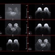
Ho and Meade imaged a variety of nanodiamond samples, including nanodiamonds decorated with various concentrations of gadolinium(III), undecorated nanodiamonds, and water. The intense signal of the gadolinium(III)-nanodiamond complex was brightest when the gadolinium(III) level was highest.
Currently, the researchers are exploring the preclinical application of the MRI contrast agent-nanodiamond hybrid in various animal models, as well as continuing studies of the structure of the gadolinium(III)-nanodiamond complex to learn how it increases relaxivity.
Copyright © 2010 AuntMinnie.com


.fFmgij6Hin.png?auto=compress%2Cformat&fit=crop&h=100&q=70&w=100)

.fFmgij6Hin.png?auto=compress%2Cformat&fit=crop&h=167&q=70&w=250)


