Researchers are using MRI to see how two regions of the brain respond to playing video games, with the hope of using that functional information to craft training and rehabilitative exercises for both healthy individuals and neurologically impaired patients.
The study, published online January 20 in the journal Cerebral Cortex, focused on the striatum and hippocampus regions of the brain and measured volume changes in each area as a result of playing a video game.
"Video games are being marketed and used in a lot of different contexts to enhance cognitive function," said lead author Kirk Erickson, Ph.D., assistant professor in the department of psychology at the University of Pittsburgh. "The basic idea is that you play the video game, you get better at the video game, and it will transfer to better managing your checkbook, driving, or other everyday activities."
Even though previous studies have found that playing video games can help enhance one's performance of cognitive tasks, Erickson maintained that there remains limited knowledge of how to use video games to benefit training, education, and rehabilitation.
"Can we use neuroimaging and MRI techniques to examine individual variability to predict who might be more influenced by these video games and who might be more easily trained?" he said.
Brain images
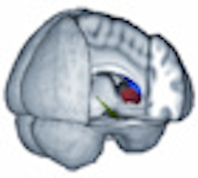
Specifically, researchers analyzed reactions in the brain's striatum and hippocampus, which are used in learning, memory, and motivation. The hippocampus is involved in more explicit types of memory functions, such as remembering a name, a recent activity, or a grocery list. The striatum remembers motor skills, such as riding a bicycle or throwing a ball, and is involved in learning other skills, such as switching tasks or multitasking.
"MRI techniques have provided us with a glimpse into the morphology and functioning of the brain in living human subjects," Erickson said. "This has been a very useful tool to examine with very high spatial resolution the details of the brain and then relate that information to these more complex tasks and everyday types of activities."
 |
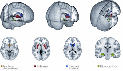
| The images show 3D and 2D displays of brain regions examined in the study -- the nucleus accumbens (orange), putamen (red), caudate nucleus (blue), and hippocampus (green). Image courtesy of Kirk Erickson, Ph.D., University of Pittsburgh. |
Participants in the study ranged from 18 to 28 years of age and were selected, in part, based on the fact that they had played video games less than three hours a week during the two years prior to the study. Twenty-six of the 36 subjects were female.
"We didn't want to take people with a lot of gaming experience, because that would have biased our results," Erickson said. "Probably not surprisingly, it is much more difficult to find males who don't have a lot of gaming experience. In future studies, we are hoping to bring that up to an even male-female ratio."
The researchers randomly assigned fixed or variable training tasks to two groups of 18 participants each, with six males in the variable task group and four males in the fixed task group.
The Space Fortress
Subjects received instructions on how to play a game called Space Fortress and were scanned on a 3-tesla MRI system (Magnetom Allegra, Siemens Healthcare, Malvern, PA) prior to two 10-hour training sessions. To assure reliability of the segmentation algorithm for the brain images, the researchers performed an additional MRI scan on all participants two weeks after the training program, using the same segmentation algorithm.
With the help of the high-resolution MR brain images, the researchers segmented different regions of the brain to isolate the striatum and hippocampus for each person. "It allowed us to calculate the volume of each structure ... and examine if we could predict their performance improvements on the video game by simply examining the volume of these structures," Erickson said.
In the analysis, the researchers found that the striatum was more related to learning how to play video games than the hippocampus. In addition, the volume of the "dorsal striatum positively predicted performance improvements for those individuals trained with strategies promoting cognitive flexibility, whereas the volume of the ventral striatum did not," the authors wrote.
Striatum variables
During early learning stages, both the volume of the ventral striatum and the volume of the dorsal striatum positively predicted performance improvements, the study noted. "These findings suggest that individual structural differences in the striatum are effective predictors of procedural learning and cognitive flexibility and are sensitive indicators of ventral-to-dorsal differences in striatal recruitment during learning."
As for the brain changes among females and males, researchers found no difference, despite the disparity in numbers. "We can be reasonably certain that our results do not reflect differential learning rates and brain volume between the genders," they stated.
The group noted, however, that the "skewed gender distribution with only 36 participants may limit the generalization of our findings and may have also affected the power to detect gender differences, if they exist. Such questions would be better addressed with larger sample sizes and an equal proportion of males and females."
Practical applications
How the information could apply to training procedures or clinical practice remains in question. "The hope is to use MRI technology to isolate the structures that a particular training technique wants to tap into and examine the individual differences before training begins," Erickson said. "Essentially, we may be able to tailor interventions or rehabilitation techniques based on the existing volumetric differences of individuals."
He and his colleagues currently are continuing their research with a larger investigation into different types of video games and training techniques and with longer training periods. They also will use different MR imaging sequences and neuroimaging techniques to view brain function beyond volume and morphology to see how function may change with various amounts of training.
By Wayne Forrest
AuntMinnie.com staff writer
February 9, 2010
Related Reading
MRI shows that exercise may increase brain volume, February 1, 2010
Neuroimaging provides clearer picture of brain dysfunction in schizophrenia, August 5, 2009
fMRI helps find schizophrenia symptoms, January 20, 2009
MRI reveals patterns of neurological abnormalities in schizophrenia, April 4, 2006
DTI scans track schizophrenia-like changes in brains of teen marijuana users, December 1, 2005
Copyright © 2010 AuntMinnie.com


