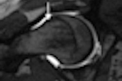
Researchers at New York University (NYU) have developed a way to use MRI to examine sodium ions in cartilage and view the development of osteoarthritis in knee joints.
The research, which appears in the Journal of Magnetic Resonance, may provide a noninvasive method to diagnose osteoarthritis in its very early stages and help to calculate other, less direct measures of cartilage assessments.
The concentration of sodium ions is known to reveal the location of glycosaminoglycans (GAGs) in cartilage tissues. GAGs are molecules that serve as the building blocks of cartilage and are involved in numerous vital functions in the human body.
GAG loss in cartilage typically marks the onset of osteoarthritis and intervertebral disk degeneration.
However, the existing techniques for GAG monitoring, which use conventional MRI, are limited in that they cannot directly map GAG concentrations, or they require contrast agents to reveal the location of these concentrations.
To better target the location of sodium concentrations, NYU researchers focused on the differences in the magnetic behavior of sodium ions residing in different tissues. By exploiting the properties of sodium ions in different environments, the team was able to develop a new method to isolate two pools of sodium ions.
As a result, they were able to obtain images in which the sodium signals appear exclusively from regions with cartilage tissue and potentially provide a way to diagnose osteoarthritis in its very early stages.
Related Reading
3T MRI aids success of surgical meniscus repairs, July 23, 2010
MRI can identify osteoarthritis risk factors 10 years early, June 30, 2010
Software saves time by segmenting meniscus in knee MR images, February 1, 2010
Sodium MRI at 3 tesla detects early signs of osteoarthritis, February 5, 2009
U.K. study finds direct access to knee MRI is cost-effective, April 3, 2008
Copyright © 2010 AuntMinnie.com


