Wednesday, December 1 | 11:30 a.m.-11:40 a.m. | SSK10-07 | Room E451B
In this scientific session, researchers will discuss how high-resolution 3D MRI can help determine the magnitude and local distribution of load-dependent cartilage deformation in the different compartments of the knee joint.According to the group from Ludwig Maximilian University (LMU) Munich Hospital in Germany, their findings may provide a base for models to calculate stress patterns and help to develop possible preventive measures for protecting workers and cartilage graft engineering.
MR images of the knee at 3 tesla were acquired in 10 healthy volunteers before and immediately after kneeling at a 90° angle, squatting, sitting on their heels for 10 minutes, and again after 90 minutes of rest after exercise.
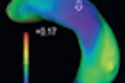
At each time point, the subjects' patellar, femoral, and tibial cartilage were segmented and reconstructed into 3D images. The researchers also calculated volumetric parameters to include volume, bone-cartilage-interface, mean thickness, and voxel-based distribution of local cartilage thickness and thickness difference plots.
"MRI provides direct depiction of articular cartilage without exposing the patient to radiation and without an invasive intervention," said study co-author Annie Horng, MD. "This renders MRI a suited technique for cartilage research, especially when one wants to examine healthy volunteers, as in our case."
Reproducibility of segmentation was 1.1% for volume, 0.6% for bone-cartilage-interface, and 0.7% for mean thickness. The group found no significant change of bone-cartilage-interface after exercise and after rest.
Significant cartilage deformation was found for volume/mean thickness of patellar (4.9%/4.7%) and lateral tibial (2.5%/2.5%) cartilage after kneeling; for volume of femoral (2.1%), mean thickness of patellar (2.2%), and volume/mean thickness of lateral tibial (2.2%/1.8%) cartilage after squatting; and for volume/mean thickness of patellar (3.1%/2.6%) and femoral (2.2%/1.4%) cartilage after volunteers sat on their heels.
"The findings show initial direct deformation areas in the patellar cartilage with additional quantitative evaluation for postures, which are assumed to be potentially harmful for the cartilage when the individual has been overexposed for a respective time frame or duration," Horng said. "Clinically, this information might be useful in improving personalized treatment, such as customized physiotherapy or medical equipment for optimizing individual joint biomechanics or maybe facilitate the choice of an appropriate cartilage replacement technique with adequate loading requirements for the diseased cartilage."
Horng and colleagues are extending the research to all cartilage plates of the knee joint to investigate the complete cartilage deformation pattern. "It would be especially interesting to see whether particular postures or exercises result in a specific deformation pattern," she added.


.fFmgij6Hin.png?auto=compress%2Cformat&fit=crop&h=100&q=70&w=100)


.fFmgij6Hin.png?auto=compress%2Cformat&fit=crop&h=167&q=70&w=250)


