Monday, November 28 | 11:30 a.m.-11:40 a.m. | SSC06-07 | Room E353B
MRI can accurately estimate the dominant tumor volume and provides greater accuracy in distinguishing tumors larger than 0.5 cm compared with serum prostate-specific antigen (PSA) and patient age, according to a new study from the U.S. National Cancer Institute (NCI).Researchers also concluded that dominant tumor volume can be used in treatment planning, specifically to identify tumor margins for focal therapy and to select candidates for active surveillance, the researchers also concluded.
"Prostate MRI is increasingly employed to help guide the biopsy and treatment of prostate cancer," said presenter Dr. Baris Turkbey from the NCI's Center for Cancer Research. "In particular, there is growing interest in minimally invasive focal therapies to avoid well-known morbidities, such as urinary incontinence and erectile dysfunction, associated with whole-gland therapy."
Turkbey and colleagues addressed the issue by investigating whether MR-derived tumor volumes predict the dominant tumor volume as determined by histopathology.
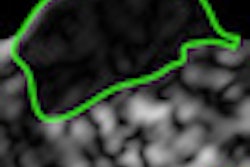
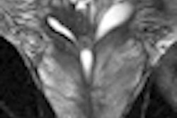
The study evaluated 135 patients with a mean age of 59.3 years who had undergone a 3-tesla MRI exam and subsequent radical prostatectomy. Dominant tumor volume was determined prospectively on T2-weighted MRI and independently estimated by measuring volumes on histopathology slides.
The results indicate a positive correlation between dominant tumor volume and MR-derived tumor volumes, and they establish that MRI is accurate in predicting the volume of tumors larger than 0.5 cm3.
"MRI appears to be an accurate method of identifying the margins of dominant prostate cancers and is suitable for guiding focal therapy," Turkbey said. "In the near future, the accuracy of MRI for determining tumor margins may be pivotal in guiding minimally invasive treatments such as image-guided focal therapy."


