Monday, November 28 | 11:50 a.m.-12:00 p.m. | SSC12-09 | Room N226
With the help of functional MRI (fMRI), Swiss researchers have found evidence of how patients can maintain their language function despite a tumor in the corresponding area of the brain.The study results suggest that language function is shifted to a different region of the brain to compensate for the deficiency.
The researchers enrolled 57 right-handed patients (32 males; mean age, 44.3 years) with brain tumors localized in the left hemisphere of the brain, and compared them with 14 right-handed healthy volunteers (7 males; mean age, 27 years).
"Functional MRI can be used to detect areas in the brain which are responsible for motor, sensory, and speech function," said presenter Sasan Partovi, a medical student at University Hospital Basel and University Hospital Bruderholz. "In our study, it was interesting to investigate with fMRI whether patients with brain tumors reveal language activation in other sites in the brain in comparison to healthy volunteers."
Each participant received a standardized whole-head blood oxygen level-dependent (BOLD) fMRI exam of language function at 1.5 tesla, using visually triggered sentence generation for determining word generation.
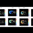
The functional MR images revealed "significant alterations" in fMRI language lateralization in patients with brain tumors, supporting the hypothesis that brain tumors affecting language-relevant areas lead to cortical reorganization and a shift of the dominant hemisphere to the contralateral side.
"This is interesting to understand, for instance, why patients with brain tumors infiltrating the language areas could nevertheless have no symptoms of language deficit," Partovi added. "Maybe the brain adapts to this new situation and starts to recruit other areas for performing the language function."
The study's findings could benefit both patients and surgeons during radical resection or neurosurgical intervention for a tumor located close to or in the language area of the brain. "Postoperatively, language deficits may result," Partovi said. "To prevent this, a language fMRI prior to surgery can be performed to determine exactly the individual language localization and lateralization."
Based on the data and the type of tumor, the neurosurgeon can plan the appropriate therapy individually for each patient, and postoperative language deficits might be prevented, Partovi added.


.fFmgij6Hin.png?auto=compress%2Cformat&fit=crop&h=100&q=70&w=100)


.fFmgij6Hin.png?auto=compress%2Cformat&fit=crop&h=167&q=70&w=250)


