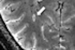
Researchers at the University of Alberta in Canada are using 4.7-tesla MRI to track the progression of multiple sclerosis (MS) in people with the disease based on increasing levels of iron found in their brain tissue.
The study of 22 MS patients and 22 subjects with no signs of MS found increased iron levels in gray-matter areas of the brain that are responsible for relaying messages. The discovery suggests there is a problem with MS patients' control system for iron. Too much iron can be toxic to brain cells, and high levels of iron in the brain have been associated with various neurodegenerative diseases.
Co-principal investigators Alan Wilman, PhD, and Dr. Gregg Blevins want to use the iron-based MRI technique to give physicians a new way to measure the effectiveness of treatments for patients with MS by watching iron levels.
The approach could open the possibility of using iron as a biomarker to assess and monitor MS over time.
The study was funded by the Canadian Institutes of Health Research, the Natural Sciences and Engineering Research Council of Canada, the Multiple Sclerosis Society of Canada, and the University of Alberta Hospital Foundation.


.fFmgij6Hin.png?auto=compress%2Cformat&fit=crop&h=100&q=70&w=100)


.fFmgij6Hin.png?auto=compress%2Cformat&fit=crop&h=167&q=70&w=250)


