Dear MRI Insider,
This issue of the MRI Insider offers an exclusive first look at how researchers are using several 7-tesla MRI sequences to visualize and characterize the substructure of multiple sclerosis (MS) lesions that cannot be seen on 1.5- or 3-tesla MRI.
Preliminary results from researchers at the University of Chicago Medical Center offer some hope of bringing 7-tesla MRI into routine clinical practice, potentially improving early diagnosis and treatment assessment for MS. Click here to read more about their progress and to see the various MR images.
The MRI Digital Community was rocked earlier this month when federal authorities arrested Chinese researchers for allegedly acquiring research money from the U.S. government and then sharing technological information with a Chinese company and a government-sponsored research institute in China. The May 20 arrests came after NYU Langone Medical Center, where the three researchers worked, conducted its own internal probe earlier this year, disclosing its findings to federal law enforcement officials.
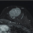
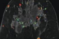
In clinical research, computer-aided detection (CAD) technology was found to improve sensitivity in breast MRI for both experienced and novice readers, but it did not significantly advance overall accuracy in a study from Washington state. There also were no significant differences in the overall time it took the 11 novice and nine experienced radiologists to interpret the breast MR images with or without CAD.
From Germany comes a study that compared the accuracy of simultaneous whole-body PET/MR, PET/CT, and retrospectively fused PET and MR images of the abdomen and pelvis. The researchers from Eberhard Karls University concluded that simultaneous PET/MRI acquisition improved the alignment of abdominal organs between PET and MRI by 51%, on average, and increased the alignment between morphological images and PET in the bladder by almost 50%.
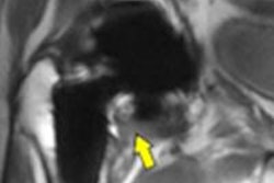
In addition, 3D volumetric MR images of cervical disease yield a bounty of critical information, enabling precise planning of brachytherapy for residual tumors, according to researchers from St. Joseph's Hospital and Medical Center in Phoenix. These 3D volumetric images, reconstructed from an isotropic 3D T2-weighted fast spin-echo sequence, facilitate excellent multiplanar tumor visualization, quantitative tumor burden assessment, and precise individual temporal response analysis for planning brachytherapy.
Be sure to stay in touch with the MRI Digital Community on a daily basis to view the latest news and novel research from around the world.

.fFmgij6Hin.png?auto=compress%2Cformat&fit=crop&h=100&q=70&w=100)



.fFmgij6Hin.png?auto=compress%2Cformat&fit=crop&h=167&q=70&w=250)
