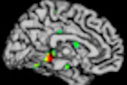
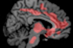
MRI may be helpful in predicting early cognitive decline by measuring changes in the fornix area of the brain, according to a study published online September 9 in JAMA Neurology.
Researchers from the University of California, Davis (UCD) found that white-matter loss in the fornix may be associated with cognitive decline in healthy elderly individuals and could help predict early clinical deterioration.
While atrophy in the hippocampus is seen in the later stages of cognitive decline, changes to the fornix and other regions of the brain structurally connected to the hippocampus are still being explored.
Lead author Evan Fletcher, PhD, and colleagues evaluated 102 cognitively normal patients (average age, 73 years) using MRI and other methods during visits over four years. The group found that changes in fornix white-matter volume and axial diffusivity were "highly significant predictors" of cognitive decline.
The study may be among the first to establish fornix degeneration as a predictor of cognitive decline among healthy elderly individuals, according to the authors.


