Thursday, December 5 | 11:40 a.m.-11:50 a.m. | SSQ15-08 | Room N229
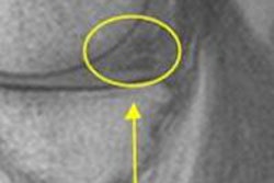
Using MR angiography (MRA), researchers from NewYork-Presbyterian/Weill Cornell Medical Center believe they have found a strong association between ischemic events and intraplaque hemorrhage.If the findings are confirmed, they believe that regular imaging of intraplaque hemorrhage on neck MRA studies could be used to determine the risk of future stroke and complement measures of luminal diameter stenosis.
Carotid plaque imaging has previously relied on high-resolution imaging with dedicated surface coils or certain MRI sequences to measure stenosis, according to radiology resident and presenter Dr. Hediyeh Baradaran and colleagues.
Recent studies suggest that 3D time-of-flight (TOF) imaging can accurately predict intraplaque hemorrhage compared to histopathology, they noted.
For their study, Baradaran and colleagues evaluated MRA neck exams performed between August 2009 and August 2012. Patients were included if they had carotid artery stenosis of 70% to 99% as seen on noncontrast 3D TOF MRA and had a history of stroke or transient ischemic attack (TIA) and other vascular risk factors.
The final study cohort consisted of 51 subjects (53 carotid arteries); intraplaque hemorrhage was found in 24 carotid arteries. Of those patients with intraplaque hemorrhage, 10 previously had suffered a stroke and five had TIAs. Of the subjects with negative results, three previously had strokes and one had a TIA.
Based on the results, Baradaran and colleagues concluded that reporting intraplaque hemorrhage on neck MRA studies could be helpful as a risk stratification tool.


