VIENNA - Ground-breaking applications of functional imaging techniques can help radiologists play their part in the fight against the emerging obesity pandemic. In particular, MR elastography is showing promise in the early detection of disease, attendees were told at Saturday's State-of-the-Art Symposium at ECR 2014.
Obesity-related illness worldwide is having a huge direct impact on healthcare costs, and indirectly affects individual loss of productivity and its related socioeconomic problems as many obese patients are deemed unfit to work. It presents an obstacle to the population's physical, mental, and financial well-being that healthcare providers can't afford to ignore.
One such associated disease is nonalcoholic fatty liver disease (NAFLD), which ranges from relatively benign, simple steatosis -- fatty accumulation in less than 5% of hepatocytes -- to a more severe condition called nonalcoholic steatohepatitis (NASH), in which there is fatty accumulation plus lobular inflammation, hepatocyte ballooning, and perisinusoidal fibrosis, and which may also lead to cirrhosis, according to Dr. Valérie Vilgrain, the chair of imaging at Beaujon Hospital in Paris.
Importantly, very recent data suggest that the first cause of hepatocellular carcinoma (HCC), the primary liver cancer, is not due any more to alcohol intake or viral hepatitis but to a metabolic syndrome in which obesity plays an important role. Furthermore, an increasing incidence of HCC, including those found incidentally, and benign liver tumors linked to obesity constituted a challenge for radiology and oncology departments. Nevertheless, NAFLD patient surveillance using imaging techniques would help to detect HCC and hepatocellular adenomas.
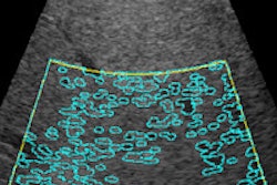
"Now our task as radiologists is to diagnose liver steatosis and quantify it, which is easy," she said, pointing to diagnosis through a number of modalities, including ultrasound, CT, and MR spectroscopy, and the lesser known controlled attenuated parameter (CAP), a measure of ultrasound attenuation on the fibroscan that correlated with pathological grades of steatosis.
"It is much more difficult to identify the dangerous subtype, NASH, defined pathologically with inflammation and fibrosis, which are harder to detect on imaging," she added.
At present, the latest development in obesity imaging is the application of functional quantitative imaging to detect inflammation and fibrosis. There is growing evidence that one such new technique, functional MR elastography, can differentiate NASH among NAFLD patients. The technique determines liver stiffness, as stiffness increases in early NASH even with no fibrosis and it further increases with the severity of the disease.
Already used routinely in Vilgrain's hospital and in dedicated centers, there is an argument for implementing MR elastography wherever obese patients are managed; not only is there a higher liver-specific mortality among NASH patients when compared with the general population, there is also a higher cardiovascular mortality, and such differentiation could influence management strategies at an earlier stage.
"We are not going to solve the problem with just morphological imaging," she commented.
Despite the difficulties generally associated with imaging obese patients, MRI might prove the most accommodating, being less subject to weight restrictions and body size -- although these were still a consideration, even with the bigger bores available today. Imaging larger patients therefore remains a challenge, despite the relative simplicity of many techniques such as conventional ultrasound. Because the liver interacts with visceral fat, which in turn interacts with the cardiovascular system and other body systems, detailed assessment and quantification of the subcutaneous and visceral fat using MR characteristics could differentiate patient prognosis, said Vilgrain, hinting at future research.
MRI also played an important role in monitoring patients after restrictive gastric bariatric therapy. Changes in fat fraction MR correlate strongly with body mass index (BMI), waist circumference and other measurements.
Detecting postsurgical complications is an important task today, and Dr. Marco Renga, a radiologist and researcher at the Sapienza Hospital in Latina, Italy, aimed to educate delegates about what to look for in the case of complications following bariatric surgery, such as extraluminal leak or communication of the gastric pouch with the excluded stomach.
Dr. Rachel Batterham, the medical and surgical lead for obesity services at University of the City of London Hospitals, was keen to underline the staggering statistics about obesity. The condition has tripled across the European countries since the 1980s, with 52% of the region's adult population now termed clinically overweight and 17% obese, meaning those with a BMI above 30.
The direct costs to healthcare services of obesity, such as related diabetes and the indirect costs to the social welfare system stemming from obesity-related illness preventing employment, are also compounded with financial costs to individuals who buy expensive but ineffective diet products and join gyms.
"Health economics is a big issue, because there is only so much money to go round," she said. "It's difficult to put a figure on this but it's estimated that 460 billion euros a year is spent in Europe on obesity."
Providing food for thought on why we were prone to obesity, Batterham explained that in prehistory, those that could eat the most and put on weight would be the ones that would survive a famine.
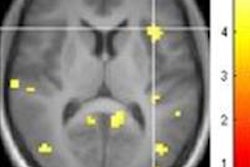
"Our genes that regulate body weight have developed in a completely different environment from the one we live in now, so we don't have any mechanisms to defend against putting on weight," she noted. "Multiple brain systems make eating rewarding."
This was one reason why surgery was a more effective means to help patients lose weight to regain health than diet and exercise alone, she said.
Originally published in ECR Today on 9 March 2014.
Copyright © 2014 European Society of Radiology


