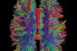
VIENNA - Emerging functional MRI, diffusion tensor imaging, and diffusion kurtosis imaging can provide a better understanding of the microstructure and function of the brain, and help to elucidate some of the mechanisms involved in neuroplasticity, explained Dr. Paul Parizel, head of the department of radiology at Antwerp University Hospital in Belgium, in Saturday's Wilhelm Conrad Röntgen Honorary Lecture.
Parizel began his presentation with a historical journey showing the brain's ability to fascinate people through the ages. According to Hippocrates (460-370 BC), "I hold that the brain is the most powerful organ of the human body ... wherefore I assert that the brain is the interpreter of consciousness."
During the Renaissance, dissection of the human brain was performed by Andreas Vesalius (1514-1564), a Flemish-born anatomist whose work helped to correct misconceptions dating from ancient times.
 Dr. Paul Parizel from Antwerp, Belgium.
Phrenology, which used bumps to localize function in the brain, was developed by German physician Franz Joseph Gall in 1796. It was based on the concept that the brain is the organ of the mind, which is composed of multiple distinct, innate faculties. Each faculty must have a separate seat or "organ" in the brain; the size of an organ is a measure of its power, and the development of various organs determines the shape of the brain, and hence the shape of the skull, according to Gall.
"The 'scientific' foundation of phrenology was epitomized by the American Phrenological Journal. No doubt it had a wonderful editor in chief and an extremely high impact factor!" Parizel remarked.
Nonscientists, too, have become intrigued by the brain. As the German fashion designer Karl Lagerfeld once said, "The brain is a muscle, and I'm a kind of bodybuilder."
Some neuroscientists have even postulated that there are parallels between the brain and the universe, Parizel said. The development of the universe may share common features with the growth of the Internet and social networks such as Facebook.
Parizel pointed out that the brain can modify its structure and function in response to external and internal stimuli, such as learning, meditation, injury, and hormonal changes.
Furthermore, the process of neuroplasticity (i.e., the ability of the brain to change, adapt, and reorganize neural pathways as it needs) offers hope to patients for recovery from injury and response to disease; imaging techniques can help to visualize neuroplasticity in terms of white-matter rewiring and structural modifications. For instance, changes in hormonal concentrations in women during the menstrual cycle, and with use of contraceptives, influence neuroplasticity changes in white matter fiber density and gray matter volume.
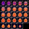
Sex hormones have an effect on brain structures and influence short-term plasticity. Coronal, axial, and sagittal slices can show the relationship between gray matter volume and estradiol concentrations in the peripheral blood of women with a natural cycle. There is a negative correlation in the anterior cingulate gyrus during the luteal phase, he elaborated.
Parizel acknowledged the input and support of his colleagues: C. Venstermans, F. De Belder, R. Salgado, L. van den Hauwe, J. Van Goethem, T. Van der Zijden, M. Voormolen, W. Van Hecke, T. De Bondt, A. Leemans, F. Deferme, and M. Geldof.
Originally published in ECR Today on 9 March 2014.
Copyright © 2014 European Society of Radiology

