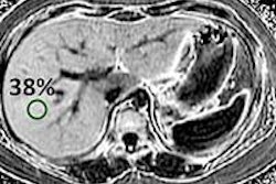
Cardiac MRI is an accurate technique for evaluating changes in the myocardium of patients with end-stage liver disease, particularly those who have been infected with hepatitis C, according to a new Egyptian study presented at ECR 2014.
Specifically, severe liver cirrhosis secondary to infection with hepatitis C causes functional and morphological changes within the myocardium that could be accurately evaluated by cardiac MRI, explained study author Dr. Noha Behairy.
There is a wide range of myocardial involvement in patients with hepatitis C and cirrhotic cardiomyopathy, said Behairy, an assistant professor in the radiology department of Cairo University, during her presentation. Cardiac failure is an important cause of death in patients undergoing liver transplantation, she added.
Egypt's high hepatitis C rates
Worldwide, hepatitis C virus infection accounts for approximately 40% of all chronic liver disease, leading to around 10,000 deaths annually, and it is the most frequent indication for liver transplants, she said. Egypt has the highest prevalence of hepatitis C in the world, ranging from 6% to 28%.
"We have a very high incidence of hepatitis C virus liver cirrhosis in Egypt, reaching an average of 13.8%," Behairy told AuntMinnie.com. "Most of those patients undergo liver transplantation."
Because cardiac failure is a common postoperative complication for liver transplant patients, the researchers wanted to find a way to select patients suitable for the procedure, especially since it is a costly operation.
Therefore, Behairy and her colleague utilized cardiac MRI to evaluate the functional and morphological myocardial changes in hepatitis C patients with end-stage liver disease listed for liver transplantation.
They included 25 patients in the study: 23 men and two women, with a mean age of 55. All of the study participants underwent routine laboratory investigations, upper gastrointestinal endoscopy, abdominal ultrasound, 2D echocardiography, and cardiac MRI on a 1.5-tesla scanner.
Balanced fast-field echo in two and four chambers and multislice short-axis images were acquired in 25 cardiac phases. Delayed enhancement was performed 10 to 15 minutes after intravenous contrast injection. The researchers analyzed the images for volumetric measurements, functional analysis, myocardial tissue characterization, and the pattern, site, and percentage of ventricular enhancement.
All of the patients showed adequate left ventricle contractility with normal wall motion and thickness, and a normal range of ejection fraction from 55% to 80% (mean, 65.5% ±11.8%). The researchers also found variable degrees of delayed myocardial enhancement in 21 of the patients, and there was a significant negative correlation between the percentage of delayed myocardial enhancement and the ejection fraction, cardiac output, cardiac index, and serum albumin levels.
Behairy told AuntMinnie.com that this is the first study to report this negative correlation, and they were particularly surprised by the negative correlation between the percentage of myocardial enhancement and serum albumin levels.
Approximately 84% of the patients showed midwall patchy and/or linear patterns of late myocardial enhancement, ranging from 2% to 52% of the myocardium, while 12% did not show any enhancement.
One limitation of the study was that patients with severe ascites experienced degraded examinations because they were unable to hold their breath properly; some study participants had to be excluded for this reason, Behairy noted.
She told AuntMinnie.com that the study's findings can be useful as a starting point for further research about the effects of hepatitis C on the heart.
"Preoperative cardiac MR may help in selecting patients suitable for liver transplantation, hence decreasing the mortality rate caused by cardiac failure," Behairy said in concluding her ECR presentation.


.fFmgij6Hin.png?auto=compress%2Cformat&fit=crop&h=100&q=70&w=100)


.fFmgij6Hin.png?auto=compress%2Cformat&fit=crop&h=167&q=70&w=250)


