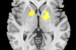
Chinese researchers have concluded that MRI of glutamate concentrations in the brain can detect ischemic lesions in the first three hours after stroke that are not seen on T1- and T2-weighted MRI.
Lead author Dr. Zhuozhi Dai of Second Affiliated Hospital of Shantou University Medical College presented the findings on Monday at the American Roentgen Ray Society (ARRS) meeting in San Diego.
Dai and colleagues imaged glutamate -- a metabolite in the brain associated with many neuropsychiatric diseases -- using an MRI technique called chemical exchange saturation transfer (CEST) to explore ischemic stroke. The study included a 7-tesla animal MRI scanner with a standard volume coil to image 12 adult male rats.
The key finding is that glutamate imaging in the first three hours after stroke showed the ischemic lesions, which were undistinguishable in T1-weighted and T2-weighted MR images.
For patients, MRI of glutamate could potentially lead to an early diagnosis and more appropriate treatment for stroke during the critical first hours, according to the researchers.


