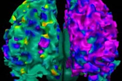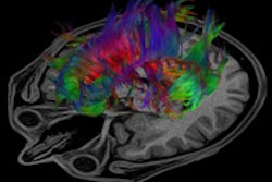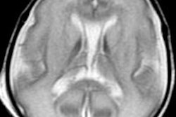
For the first time, researchers are using MRI to calculate the volume of multiple brain regions in newborns to investigate how those areas develop based on gender, gestational period, and other factors in the first 90 days of life.
The study from researchers at the University of California, San Diego (UCSD) and the University of Hawaii was published online on Monday in JAMA Neurology. Among other findings, the group determined that the normal newborn brain grows, on average, 1% each day immediately following birth, but growth slows to 0.4% per day by the time the infant reaches 3 months.
"Our findings give us a deeper understanding of the relationship between brain structure and function when both are developing rapidly during the most dynamic postnatal growth phase for the human brain," lead author Dominic Holland, PhD, said in a statement. Holland is a researcher in the department of neurosciences at UCSD School of Medicine.
Assessing brain regions in terms of growth rate, size, and asymmetry could help detect the earliest signs of neurodevelopmental disorders such as autism or perinatal brain injury.
Also, the study helps fill a void of imaging research detailing early postnatal brain development in healthy-term infants, according to Holland and colleagues.
There was a "serious gap in our knowledge of the normally maturing brain, because the earliest periods of postnatal development are the most dynamic and may have pronounced bearing on neurodevelopmental disorders, particularly those eventually manifesting as childhood-onset neuropsychiatric disorders," they wrote (JAMA Neurology, August 11, 2014).
Their longitudinal study included MRI exams of 87 healthy newborns performed between October 2007 and June 2013. The subjects included both premature and full-term infants, and there were 39 boys and 48 girls. T1- and T2-weighted MR images were acquired with 3D pulse sequences on a 3-tesla scanner (Magnetom Trio Tim, Siemens Healthcare) while the infants slept without sedation.
Brain growth
The researchers looked at 211 time points, focusing on cerebral structural development in the first three months after delivery. Regions of interest included the lateral ventricles, hippocampus, caudate, and putamen, as Holland and colleagues investigated factors such as left-right brain asymmetries, growth rates based on age, and volume according to age and gender.
They found that the newborn brain grows approximately 1% each day immediately following birth, but growth slows to 0.4% per day by 3 months of age. Overall growth was 64% in the first 90 days. As was expected, gestational age at birth significantly affected structure size for all regions of interests, except the pallidum and third ventricle. A longer gestation period was associated with larger brain size at birth.
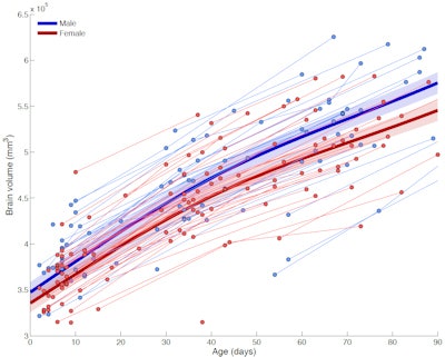 Plot shows brain volume versus age for infants during their first 90 days. Joined points represent single individuals followed over time. Boys' brains (blue) grew slightly faster than girls' brains (red). Overall, the growth rate dropped from about 1% to 0.4% per day at 90 days. Image courtesy of Dominic Holland, PhD.
For a baby born prematurely by one week, brain size was 4% to 5% smaller than what is expected for a full-term baby, the authors noted. They found that the premature infants' brains actually grew faster than full-term babies' brains; however, at 90 days after delivery, premature infants' brains were still 2% smaller.
While gender did not influence a newborn's cerebral structure at birth, at 90 days, male brains had grown more rapidly (66%) than female brains (63%). In general, the cerebellum, which is associated with motor control, grew at the fastest rate; the region more than doubled in size -- 113% growth in boys and 105% in girls -- in 90 days. The hippocampus, which is linked to memory, grew at the slowest rate, gaining only 47% in volume.
The researchers also found that many asymmetries in the brain are already established in the early postnatal period, including the right hippocampus being larger than the left. Previous studies had suggested that this occurs in the early adolescence.
"Cerebral asymmetry has been associated with asymmetric or lateralized functional specializations seen in adults, including handedness, dexterity, and language abilities," the authors wrote. "In addition, several neurological disorders are believed to be developmental and are associated with atypical brain asymmetry, particularly schizophrenia and autism. Thus, early deviant brain asymmetry ... may provide an index for individuals being at risk for a neurodevelopmental disorder.
Holland and colleagues plan to continue their efforts by applying different MRI techniques to explore the newborn brain. Future studies could also shed light on any connections between alcohol and drug use during pregnancy and infant brain volume and growth.


