Researchers from several institutions have used a 21-tesla preclinical MR spectroscopy scanner to develop a new technique for evaluating patients for stroke and possibly neurodegenerative diseases. They believe the protocol could be migrated to 3- and 7-tesla MRI scanners being used clinically.
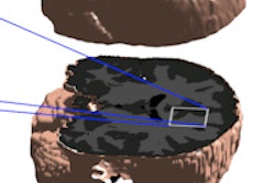
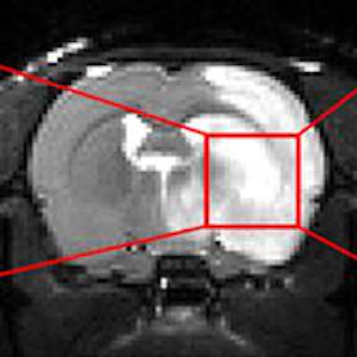
The group from the National High Magnetic Field Laboratory at Florida State University (FSU), the Champalimaud Center in Portugal, and the Weizmann Institute of Science in Israel has crafted an imaging sequence that targets specific metabolites in certain regions of the brain to determine their concentration and other information.
The key piece of technology is FSU's 900-MHz, 21.1-tesla nuclear MR magnet system, which can visualize chemical signatures of metabolites in 125-microliter volumes. The technique details metabolites using a relaxation-enhanced MR spectroscopy (MRS) approach that accentuates the metabolites with the very high magnetic field.
"The truly revolutionary part is that we can focus our attention and selectively excite certain regions of the brain by focusing on certain frequencies," said Samuel Grant, PhD, associate professor of chemical and biomedical engineering and MRI user program director. "As a result, we have more sensitivity and the ability to detect things such as metabolites and, potentially, neurotransmitter changes. Very small signal alterations give us the added benefit of looking at how [metabolites] deviate from normal levels, as with a stroke or other neurodegenerative disease that might start to evolve."
Metabolites such as choline, creatine, and lactate are small molecules that are involved in chemical reactions of every cell in a living organism. By analyzing their distribution in brain tissue, researchers can measure changes and better assess the degree of a stroke.
"We can investigate their location to potentially give us some additional information on stroke severity, the potential of the stroke outcome, and how these metabolites are interacting during the ischemic event when there is a reduction in oxygen to that brain region," added fellow FSU lab researcher Jens Rosenberg.
','dvPres', 'clsBtn', 'true' );" >

Click image to enlarge.
So far, the researchers have used the technique to image stroke rates in vivo, and they have used it ex vivo on some brain samples. The possibility of translating the technique to the human patient population in a clinical setting is actually "very high," Grant said.
"We have done most of the work at 21.1 tesla, which is seven times higher than most clinical scanners," he said. "Even at lower clinical magnetic fields, like 3 tesla and 7 tesla, we could have the ability to translate these types of techniques to benefit a larger patient population."
The challenge now, of course, is to adapt the imaging technique to more conventional 3-tesla MRI scanners. But even at higher field strengths, Grant expects patients to have little difficulty in tolerating a scan.
"In many respects, it will be very beneficial, because the big benefit from [increased] signal to noise and sensitivity allows us to reduce the scan time significantly," he said. "These types of chemical analyses now can be done feasibly in seconds or minutes -- as opposed to longer scans that are typically required -- with no real negative impact on the patient."
Technological challenges
Some technical hurdles remain to optimizing the technique on a conventional clinical MRI scanner. There will be somewhat of a reduction in sensitivity due to the lower magnet strength, but the researchers say the end results will still be helpful in evaluating stroke patients.
"One of the big issues we have is the single-voxel technique, which means we can look at one volume of the brain and investigate that very well," Grant said. "Right now we are at the 5-mm cubic voxel size. We can push that down toward 1-mm sizes, but even at 5 mm, it is very clinically relevant."
Grant and colleagues have spoken to a few clinical MRI vendors about their progress, but the technique is not quite ready for prime time. However, he asserted that MRI systems on the market today should be able to accommodate the additional feature.
"It doesn't have to be a hardware refinement by any stretch of the imagination," he said. "It basically is the implementation of the software that can handle the technique. I think it is something that can be very easily translated to the clinical side."
The FSU lab is run by the National Science Foundation. Grant said the facility wants to provide high-field technical assistance to other researchers in specialties such as physics, geochemistry, and biological and biomedical work.
"It is a good opportunity for people to come and, even before it is a clinical product, do some research on their preclinical animal models on different diseases and see if there is some benefit and utility," he added.
The research is further detailed in two recent papers, one in Nature Communications (September 17, 2014) and one in the Journal of Cerebral Blood Flow and Metabolism (September 10, 2014).


